Detection of cancer cells in body fluids

Active Publication Date: 2006-06-01
JOHN WAYNE CANCER INST
View PDF9 Cites 39 Cited by
  • Summary
  • Abstract
  • Description
  • Claims
  • Application Information

AI Technical Summary

Benefits of technology

[0007] The present invention provides new markers and new combinations of markers for detecting melanoma and carcinoma cells in body fluids. The invention also provides a quantitat

Problems solved by technology

The staging system, however, does not accurately take into account the disease progression events, particularly ongoing systemic metastasis, at the time patients are seen.
Heterogeneity of the expression of tumor genes and variable perf

Method used


Image

  • Detection of cancer cells in body fluids
  • Detection of cancer cells in body fluids
  • Detection of cancer cells in body fluids

Examples


example 1

Multimarker Quantitative Real-Time PCR Detection of Circulating Melanoma Cells in Peripheral Blood: Relation to Disease Stage in Melanoma Patients

MATERIALS AND METHODS

Melanoma Cell Lines

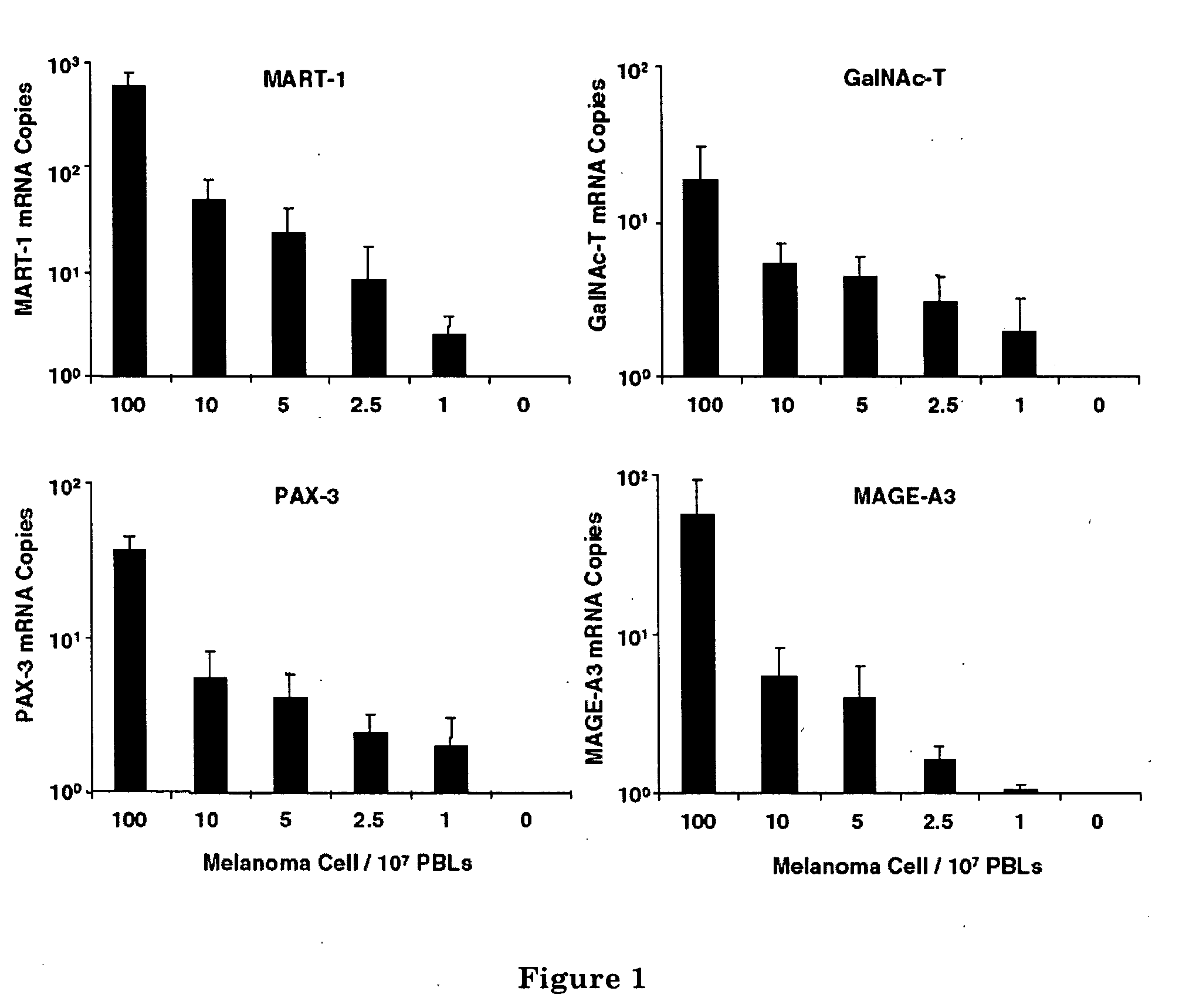
[0085] Seventeen melanoma cell lines (MA, MB, MC, MD, ME, MF, MG, MH, MI, MJ, MK, ML, MM, MN, MO, MP, and MQ) were established and characterized at the John Wayne Cancer Institute (JWCI). Cells were grown in RPMI 1640 containing 100 mL / L heat-inactivated fetal calf serum and 10 g / L penicillin / streptomycin (Gibco) in a T75-cm2 flask and were used when they reached 70%-80% confluence.
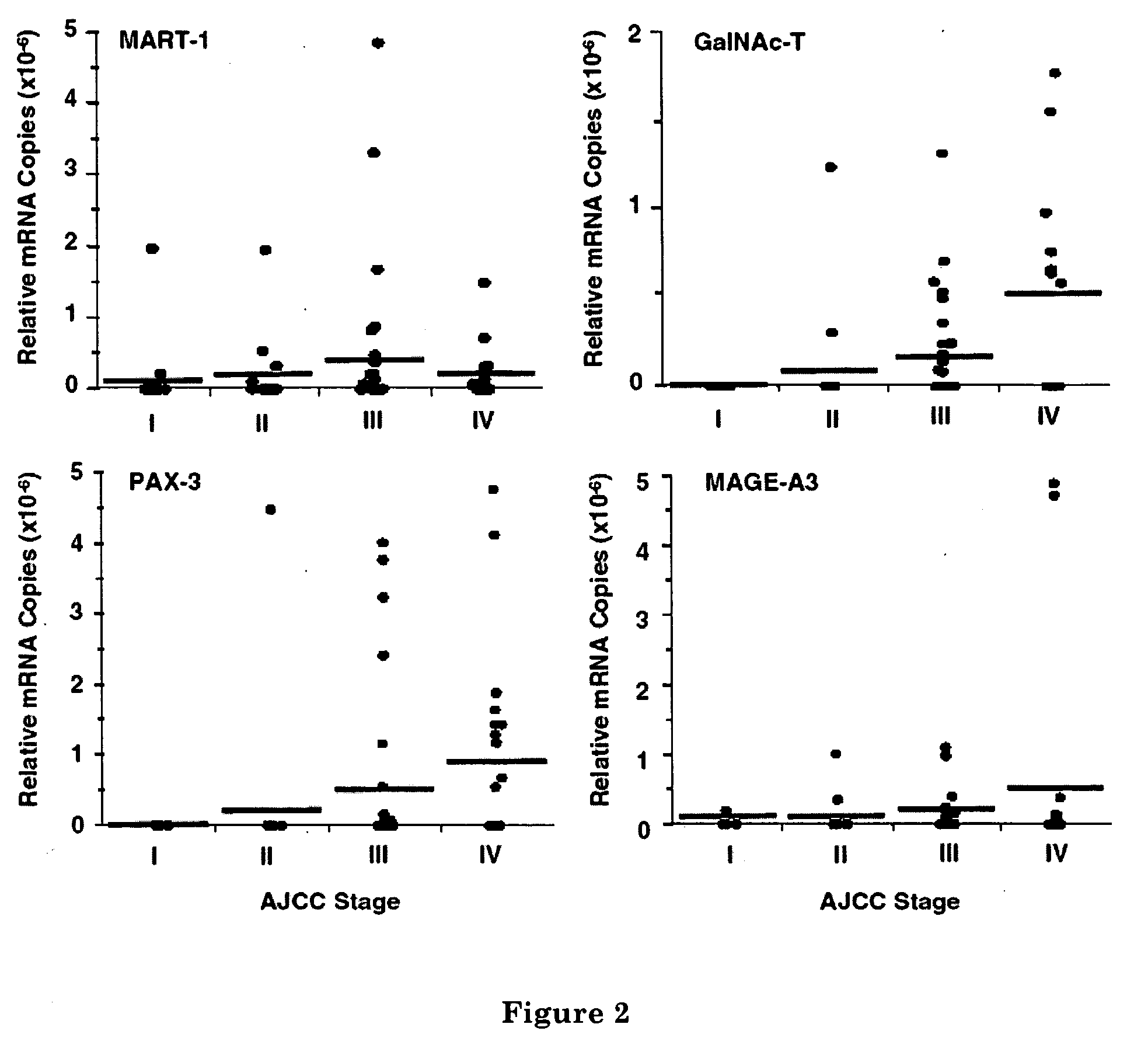
Patients

[0086] All patients enrolled in the study had documented physical and medical histories, and their AJCC stage of disease was determined and recorded at the time of blood drawing. Blood was drawn from 94 melanoma patients (20 with stage I, 20 with stage II, 32 with stage III, and 22 with stage IV disease) immediately before they received any treatment at JWCI. All patients signed consents for the use of their...

example 2

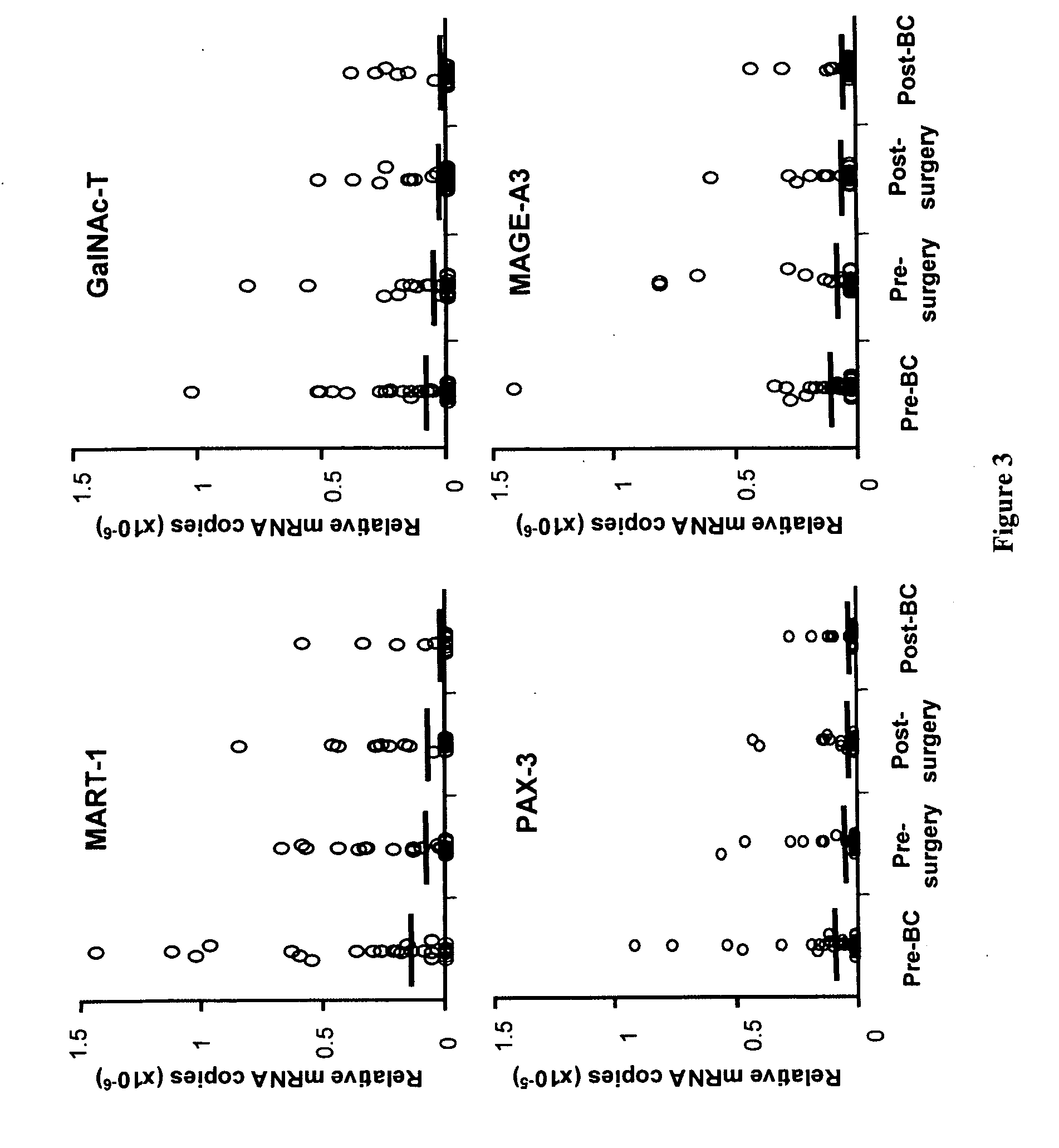
Serial Monitoring of Circulating Melanoma Cells during Neoadjuvant Biochemotherapy for Stage III Melanoma: Outcome Prediction in a Multicenter Trial


PUM

PropertyMeasurementUnit
Timeaaaaaaaaaa
Fractionaaaaaaaaaa
Fractionaaaaaaaaaa

Abstract

A method of detecting circulating melanoma or carcinoma cells in a subject. The method comprises obtaining a body fluid from a subject and detecting the expression of a panel of genes in the body fluid, wherein the expression of the panel of genes indicates the presence of circulating melanoma or carcinoma cells in the subject. Genes useful for detecting melanoma cells includes GalNAc-T, MAGE-A3, MART-1, PAX-3, and TRP-2; genes useful for detecting carcinoma cells include C-Met, MAGE-A3, Stanniocalcin-1, Stanniocalcin-2, mammaglobin, HSP27, GalNAc-T, CK20, and β-HCG. Also disclosed are kits containing agents for detecting the expression of these genes.

Description

RELATED APPLICATIONS [0001] The present application is a continuation-in-part of pending prior U.S. patent application Ser. No. 10 / 713,808 filed on Nov. 14, 2003 which claims priority to U.S. Provisional Application Ser. No. 60 / 426,216 filed on Nov. 14, 2002. The present application also claims priority to U.S. Provisional Application Ser. No. 60 / 609,634 filed on Sep. 14, 2004. The contents of U.S. patent application Ser. No. 10 / 713,808, U.S. Provisional Application Ser. No. 60 / 426,216, and U.S. Provisional Application Ser. No. 60 / 609,634 are incorporated herein by reference.FUNDING [0002] This invention was made with support in part by grants from NIH National Cancer Institute P01 Grants CA 29605, Project II, and CA 12528, Project II. Therefore, the U.S. government has certain rights.FIELD OF THE INVENTION [0003] The present invention relates generally to cancer diagnosis, prognosis, and management. In particular, the invention relates to the detection of genetic markers indicative...

Claims


Application Information

Patent Timeline
IPC IPC(8): C12Q1/68C12P19/34G01N33/53
CPCC12Q1/6886C12Q2600/112C12Q2600/118
Inventor HOON, DAVE
Owner JOHN WAYNE CANCER INST
Why Eureka
Try Eureka