Portable digital tomosynthesis imaging system and method

a tomosynthesis imaging and portable technology, applied in the field of portable tomosynthesis systems, can solve the problems of inability to move or transport fixed systems, inability to carry, and further damage,

Active Publication Date: 2008-10-02
GENERAL ELECTRIC CO
View PDF2 Cites 57 Cited by
  • Summary
  • Abstract
  • Description
  • Claims
  • Application Information

AI Technical Summary

Problems solved by technology

Although this may be a relatively straightforward requirement to satisfy in “fixed” digital radiography systems with dedicated mechanical positioners for both the source and the detector, such fixed systems cannot be readily moved or transported.
In such situations, the person needing medical care normally should not be moved because of the risk of doing further damage to the s

Method used


Image

  • Portable digital tomosynthesis imaging system and method
  • Portable digital tomosynthesis imaging system and method
  • Portable digital tomosynthesis imaging system and method

Examples


Embodiment Construction

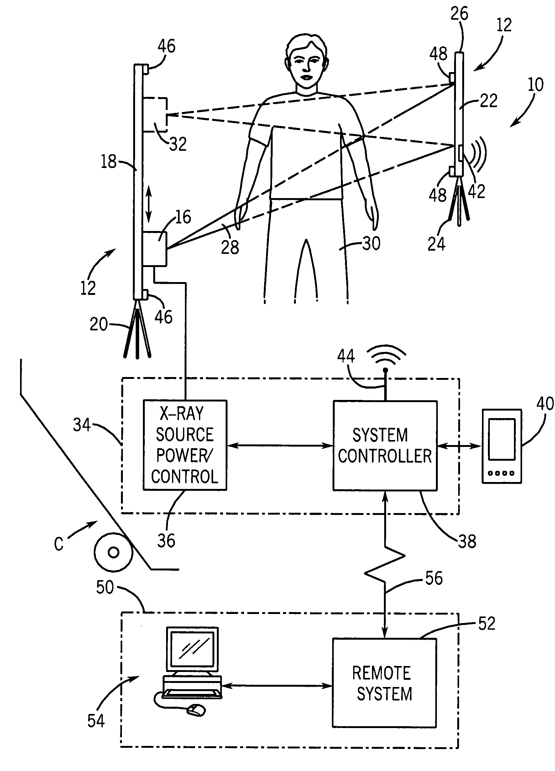
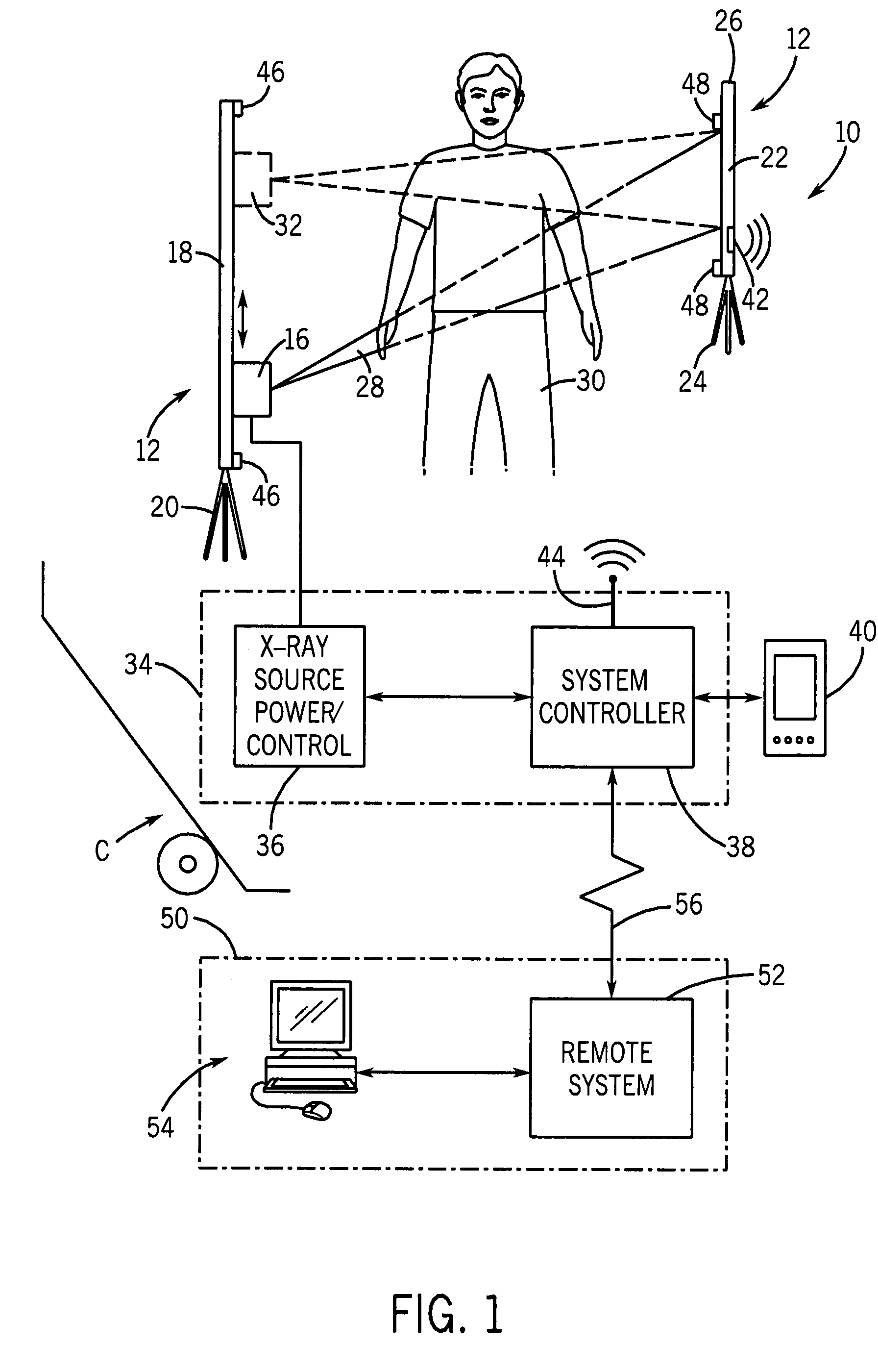
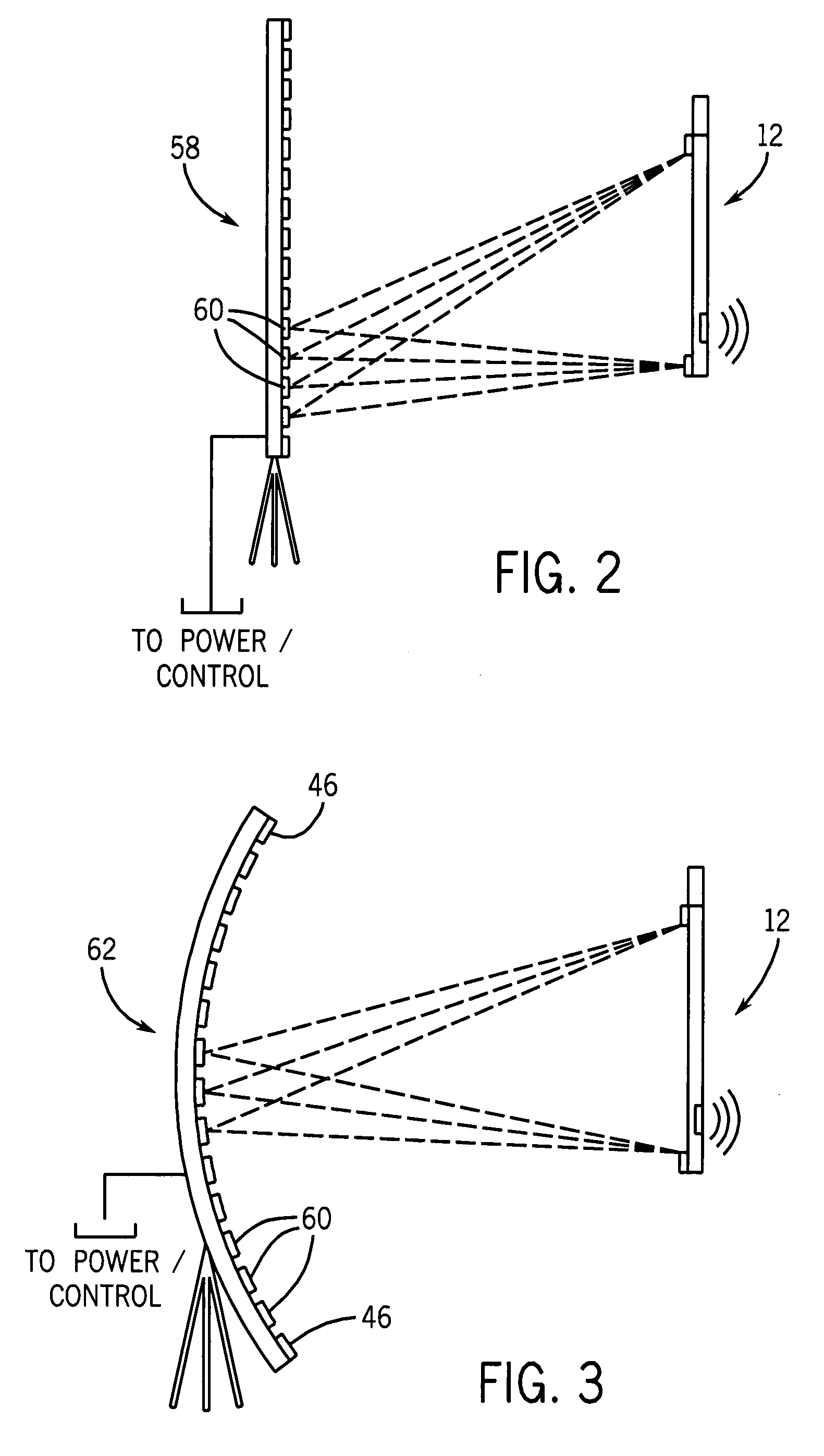
[0013]Turning now to the drawings, and referring first to FIG. 1, a portable tomosynthesis system is illustrated diagrammatically and represented generally by reference numeral 10. The system includes a portable X-ray source assembly 12 designed to cooperate with a portable detector assembly 14 to generate a series of projection images that can be used for calculating slice images through a subject of interest. In the illustrated embodiment, the source assembly 12 includes a moveable X-ray source 16 designed to slide or be drawn along a support 18. The source will typically move along a track formed in the support 18, and may move along linear or non-linear paths as discussed in greater detail below. In a presently contemplated embodiment, the source assembly 12 may include some sort of mechanical mounting structure 20, such as a tripod which can be deployed and collapsed for ease of movement and storage of the source assembly.

[0014]The detector assembly 14 may be physically separat...


PUM


Abstract

A portable tomosynthesis imaging system is disclosed that includes a portable X-ray source assembly and a portable detector assembly. The source assembly may be coupled to a portable power supply and controller such that multiple projection X-ray images may be obtained at a site that is not accessible by conventional tomosynthesis imaging systems. Image data may be transmitted from the detector by wired or wireless communication. Tomosynthesis image reconstruction may be performed locally at the portable system or remotely by the transmission of raw or filtered image data from the portable system. The portable system is particularly well-suited to field deployment, such as at accident scenes, scenes of natural or other disasters, or in confined clinical settings.

Description

BACKGROUND[0001]The present invention relates generally to digital tomosynthesis imaging systems, and more particularly to a portable tomosynthesis system suitable for use in on-sight locations, confined clinical environments, and so forth.[0002]Digital X-ray tomosynthesis is an imaging technique that enables three-dimensional imaging of a patient using a large-area digital detector typically used for conventional (single projection) radiography. In clinical tomosynthesis techniques, a source of X-ray radiation is moved between imaging positions and multiple images are made by casting an X-ray beam on a digital detector. In certain applications, multiple, distributed and static X-ray sources may be used, or movable sources may be displaced in various patterns or trajectories. In certain systems, the detector is also moved during this process. Three-dimensional data is reconstructed in the form of a number of slices through the patient anatomy, each parallel to the detector plane. To...

Claims


Application Information

Patent Timeline
IPC IPC(8): A61B6/00
CPCA61B6/025A61B6/4405A61B6/547A61B6/563
Inventor JABRI, KADRI NIZARUPPALURI, RENUKASABOL, JOHN MICHAELAVINASH, GOPAL BILIGERI
Owner GENERAL ELECTRIC CO
Why Eureka
Try Eureka