Magnetic resonance device and method for perfusion determination

a magnetic resonance and perfusion technology, applied in the field of magnetic resonance devices and perfusion determination methods, can solve the problems of low perfusion information quality of conventionally calculated mr images, prone to artifacts, etc., and achieve the effect of improving the quality of mr images depicting perfusion information

Active Publication Date: 2008-12-25
SIEMENS HEALTHCARE GMBH
View PDF22 Cites 7 Cited by
  • Summary
  • Abstract
  • Description
  • Claims
  • Application Information

AI Technical Summary

Benefits of technology

[0007]An object of the present invention is to improve the

Problems solved by technology

The acquisition of images of a relative perfusion or of images for calculation of a quantitative perfusion is therefore prone to artifacts.
The quality of the conventionally calculated MR images

Method used


Image

  • Magnetic resonance device and method for perfusion determination
  • Magnetic resonance device and method for perfusion determination
  • Magnetic resonance device and method for perfusion determination

Examples


Embodiment Construction

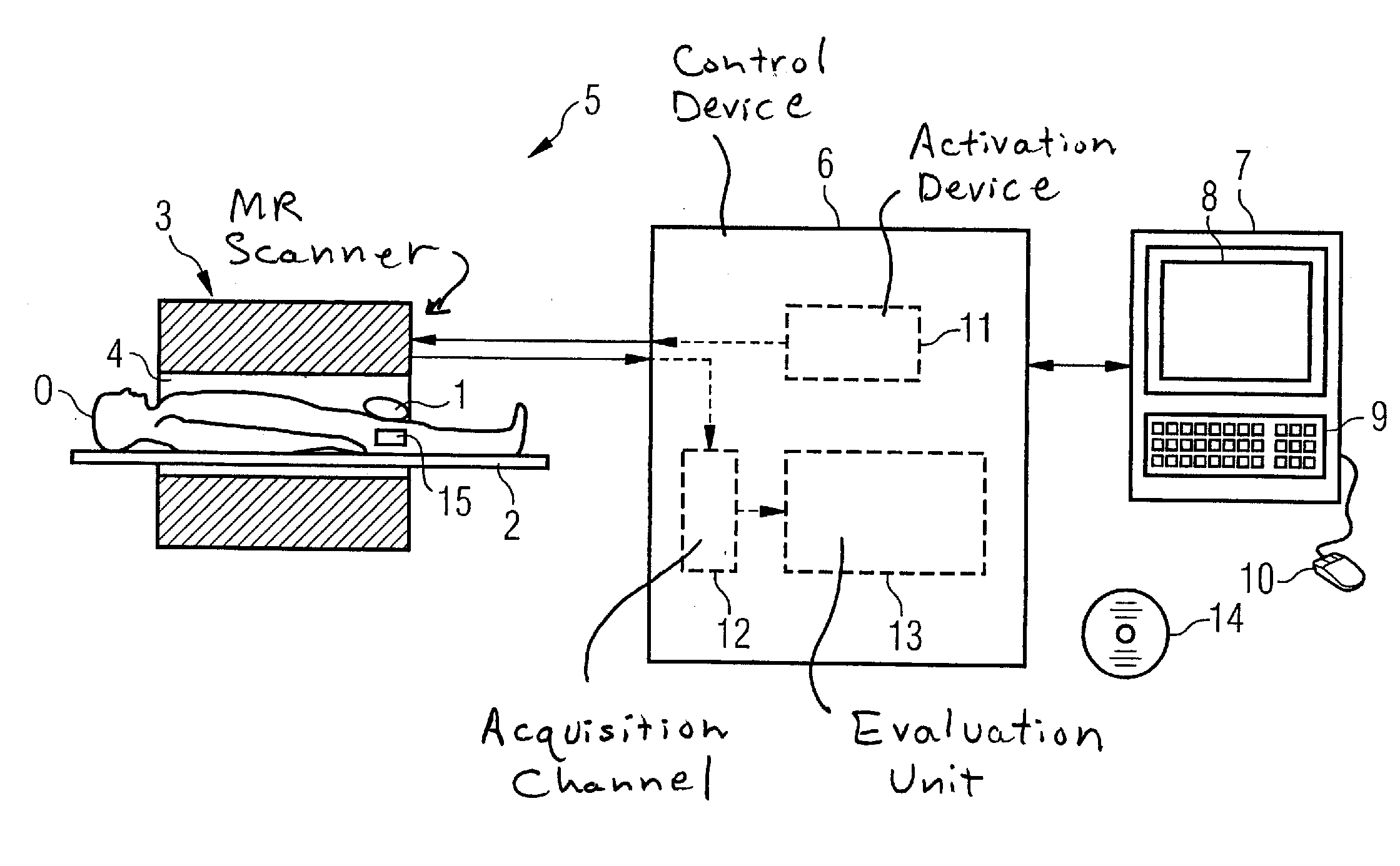
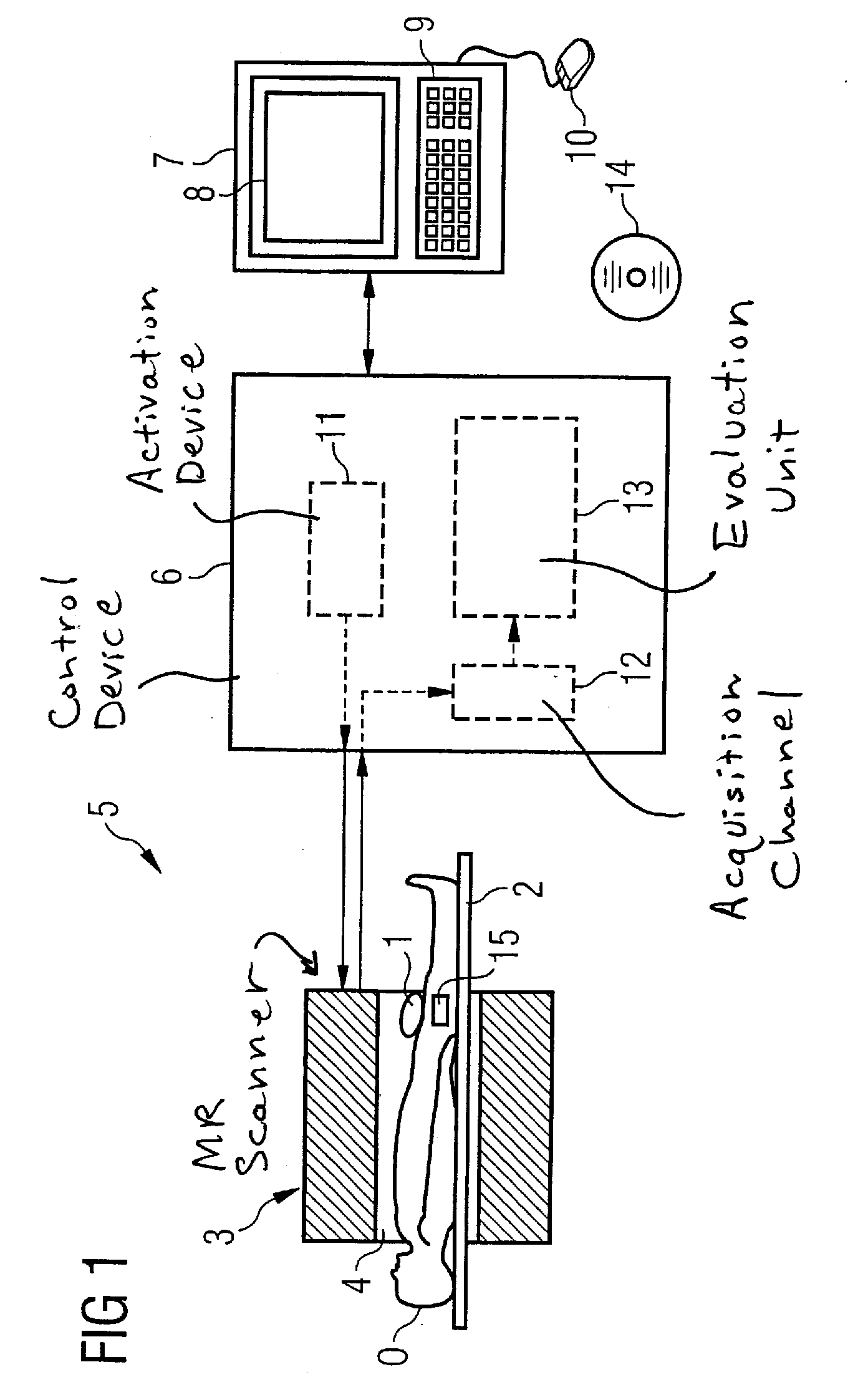
[0056]FIG. 1 shows an exemplary embodiment for a magnetic resonance system 5 with which an automatic determination of perfusion is possible. The core of this magnetic resonance system 5 is a scanner (MR data acquisition unit) 3 in which is positioned a patient O on a recumbent board 2 in an annular basic field magnet (not shown) which surrounds a measurement volume 4.

[0057]The recumbent board 2 can be displaced in the longitudinal direction, i.e. along the longitudinal axis of the scanner 3. A whole-body coil (not shown), with which radio-frequency pulses can be emitted and also received is located within the basic field magnet in the scanner 3. Moreover, the scanner 3 contains gradient coils (not shown) in order to be able to apply a magnetic field gradient in each of the three spatial directions.

[0058]The scanner 3 is controlled by a control device 6 which here is shown separate from the scanner 3. A terminal 7 that includes a screen 8, a keyboard 9 and a mouse 10 is connected to ...


PUM


Abstract

In a method and a device for automatic determination of perfusion by using a magnetic resonance system, multiple first MR data sets are thereby acquired from a volume element over time with a perfusion-sensitive imaging sequence, and multiple second MR data sets of the same volume element are acquired over time with a control imaging sequence, in particular a perfusion-insensitive imaging sequence. These first MR data sets and the second MR data sets are subjected to a statistical time series analysis in order to determine the perfusion in the volume element.

Description

BACKGROUND OF THE INVENTION[0001]1. Field of the Invention[0002]The present invention concerns a method and a device in order to automatically determine a perfusion with a magnetic resonance system. Moreover, the present invention concerns a magnetic resonance system designed with the inventive device as well as an electronically readable data medium encoded with programming instructions that cause the method to be executed by a computer.[0003]2. Description of the Prior Art[0004]MR perfusion methods (methods to determine perfusion by means of a magnetic resonance system) are used, for example, in order to measure a blood flow in various body regions, for example in the head (cerebral blood flow (CBF)). CBF is the volume of arterial blood (mL) which flows in 100 g of tissue per minute; in humans a typical value for CBF in the brain is60mL100g×min.If the density of the brain is set near to 1 g / mL, CBF in humans is 0.6mLmL×minor0.01s-1.With reference to a volume, the dimensions of the...

Claims


Application Information

Patent Timeline
IPC IPC(8): A61B5/055
CPCA61B5/0263A61B5/055G01R33/4806G01R33/56341G01R33/56366
Inventor MEYER, HEIKOPFEUFFER, JOSEF
Owner SIEMENS HEALTHCARE GMBH
Why Eureka
Try Eureka