System and method for improving workflow efficiences in reading tomosynthesis medical image data

a technology of medical image data and workflow efficiency, applied in the field of image processing, can solve the problems of a large amount of image data produced by 3d medical imaging procedures, and achieve the effects of enhancing the identification of regions, avoiding sacrificing desired detail in images, and improving workflow efficiency in reading

Active Publication Date: 2015-03-17
ICAD INC
View PDF12 Cites 65 Cited by
  • Summary
  • Abstract
  • Description
  • Claims
  • Application Information

AI Technical Summary

Benefits of technology

[0008]This invention overcomes disadvantages of the prior art by providing a system and method for improving workflow efficiencies in reading tomosynthesis medical image data that avoids sacrificing desired detail in images. The system and method generally enhances the identification of regions and / or objects of interest (ROIs), such as masses, within an acquired image by performing, based on three-dimensional (3D) data, an enhancement process to the image before it is projected into a two-dimensional (2D) format. This renders the regions / object(s) of interest more identifiable to a viewer (e.g. a diagnostician, such as a physician and / or radiologist) in the 2D-projected image as it boundaries are more-defined within the overall field.

Problems solved by technology

However, the substantial amount of image data produced by 3D medical imaging procedures presents a challenge.

Method used


Image

  • System and method for improving workflow efficiences in reading tomosynthesis medical image data
  • System and method for improving workflow efficiences in reading tomosynthesis medical image data
  • System and method for improving workflow efficiences in reading tomosynthesis medical image data

Examples


Embodiment Construction

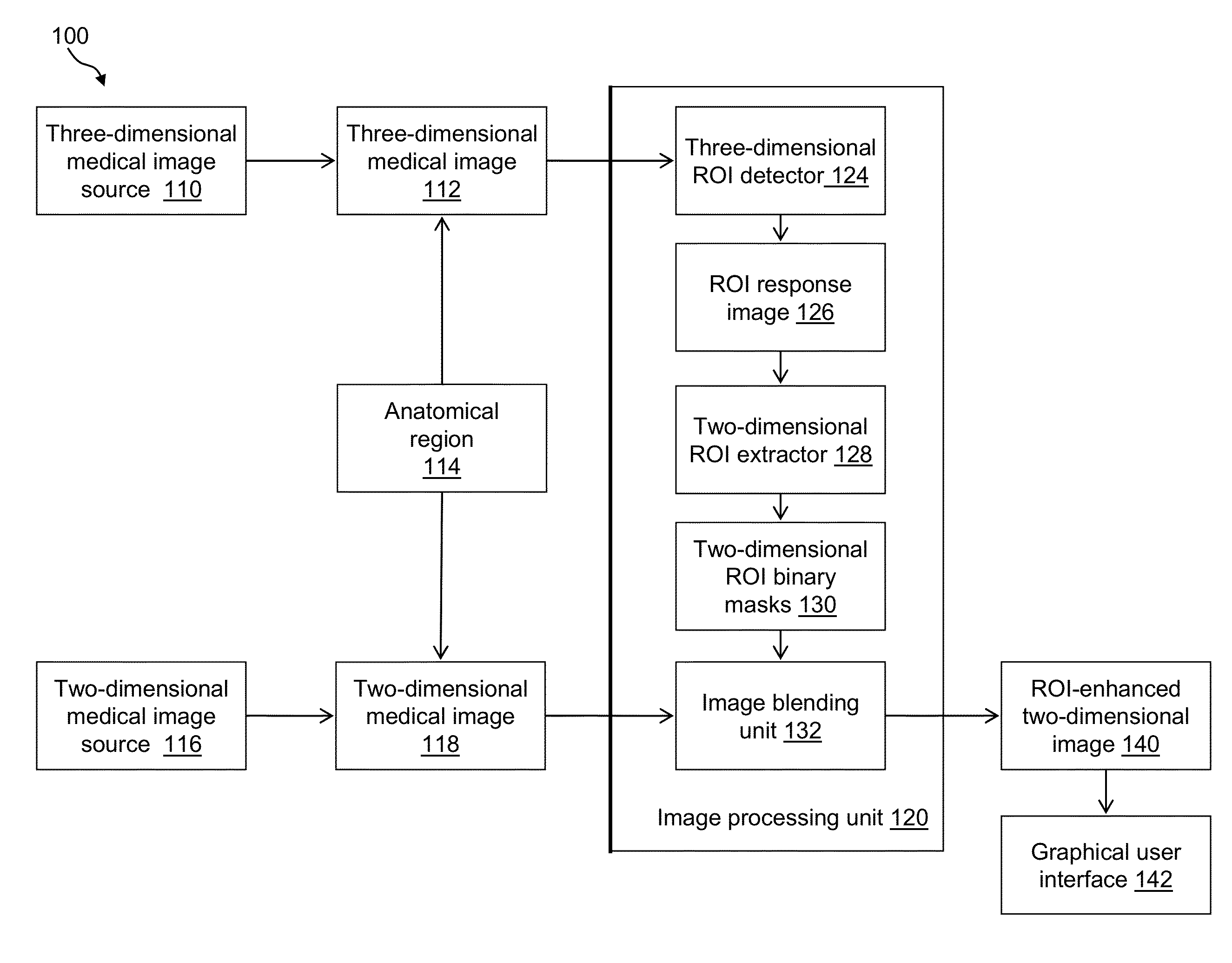
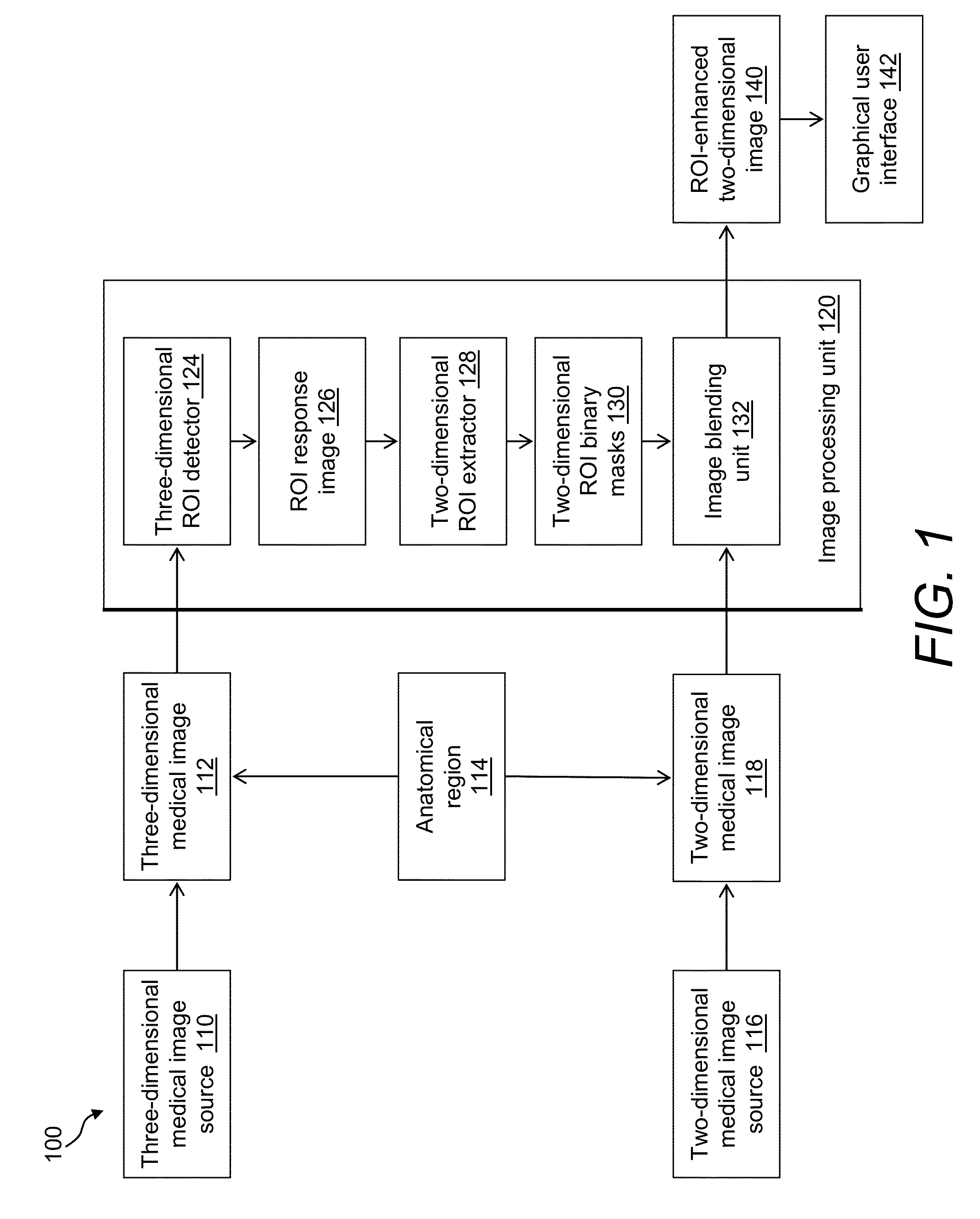
[0016]FIG. 1 is a block diagram of a medical imaging system 100 in accordance with an illustrative embodiment. The system includes a three-dimensional medical image source 110, a two-dimensional medical image source 116, and an image processing unit 120 that produces a novel, region of interest (ROI)-enhanced two-dimensional image 140 that can be the primary image read for detection and diagnosis of disease by a diagnostician. The system 100 further includes a graphical user interface (GUI) and / or display 142 for outputting the various medical image data. It should be noted that a wide range of functional components can be provided to the system, 100 in various embodiments, including various networked data-handling and storage devices, additional displays, printing devices, interfaces for portable computing devices, etc.

[0017]According to an embodiment, the three-dimensional medical image source 110 is a digital tomosynthesis imaging system such as offered by the General Electric Co...


PUM


Abstract

A system and a method are disclosed that forms a novel, synthetic, two-dimensional image of an anatomical region such as a breast. Two-dimensional regions of interest (ROIs) such as masses are extracted from three-dimensional medical image data, such as digital tomosynthesis reconstructed volumes. Using image processing technologies, the ROIs are then blended with two-dimensional image information of the anatomical region to form the synthetic, two-dimensional image. This arrangement and resulting image desirably improves the workflow of a physician reading medical image data, as the synthetic, two-dimensional image provides detail previously only seen by interrogating the three-dimensional medical image data.

Description

BACKGROUND[0001]1. Field of the Invention[0002]This application relates generally to image processing for biomedical applications. More particularly, this application relates to improving workflow efficiencies in reading medical image data.[0003]2. Description of the Related Art[0004]In the fields of medical imaging and radiology, various techniques may be employed for creating images of an anatomical region of the human body. For example, in mammography, the breast is often imaged at two fixed angles using x-rays. Physicians may review two-dimensional (2D) or planar x-ray images of the anatomical region to uncover and diagnose disease-like conditions, such as breast cancer.[0005]Numerous medical imaging procedures now employ systems and techniques that create three-dimensional (3D) or volumetric imagery of the human body. For example, significant attention has been given to tomographic imaging techniques. One such example is digital breast tomosynthesis (DBT), a relatively new imag...

Claims


Application Information

Patent Timeline
Patent Type & Authority Patents(United States)
IPC IPC(8): G06K9/00A61B6/00A61B6/02G06T7/00
CPCG06T7/0012A61B6/025A61B6/5223A61B6/502A61B6/469A61B6/466G06T5/50G06T2207/20221F04C2270/041G06T7/11G06T7/194G06T2207/10112G06T2207/30096
Inventor PERIASWAMY, SENTHILFOTIN, SERGEY
Owner ICAD INC
Why Eureka
Try Eureka