
Rapid, semi-automated method to detect respiratory virus infected cells in direct specimens
Patent Information
- Authority / Receiving Office
- US · United States
- Current Assignee / Owner
- Publication Date
- 2012-08-09
- Estimated Expiration
- Not applicable · inactive patent
Smart Images
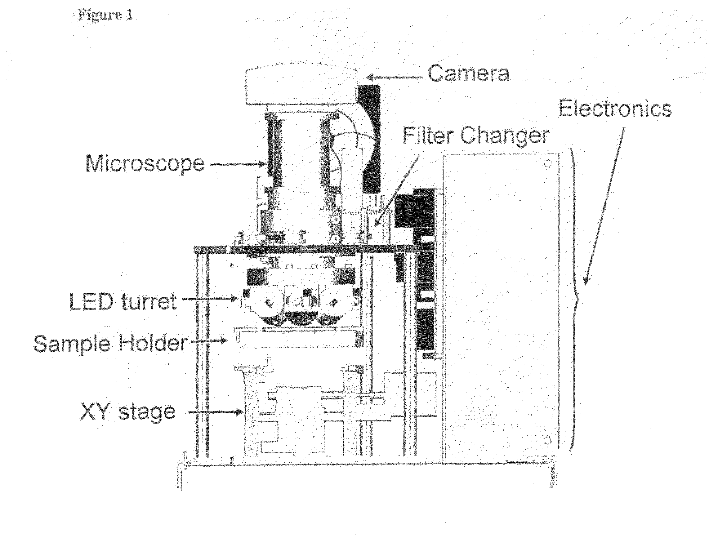
Figure 1 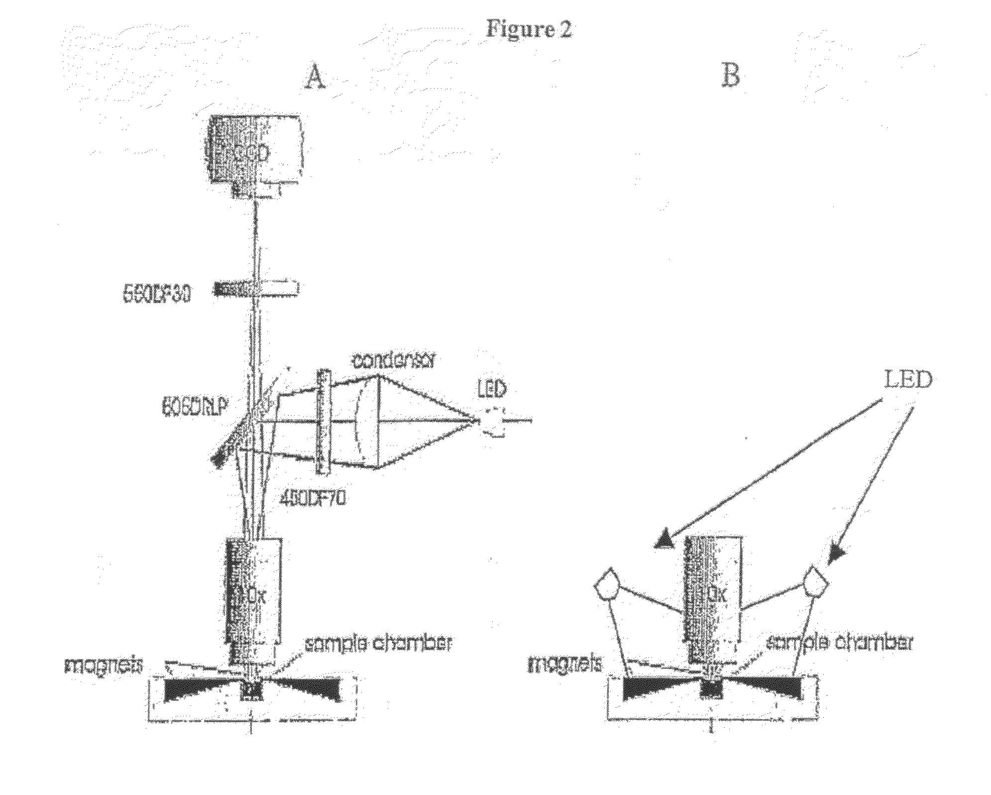
Figure 2 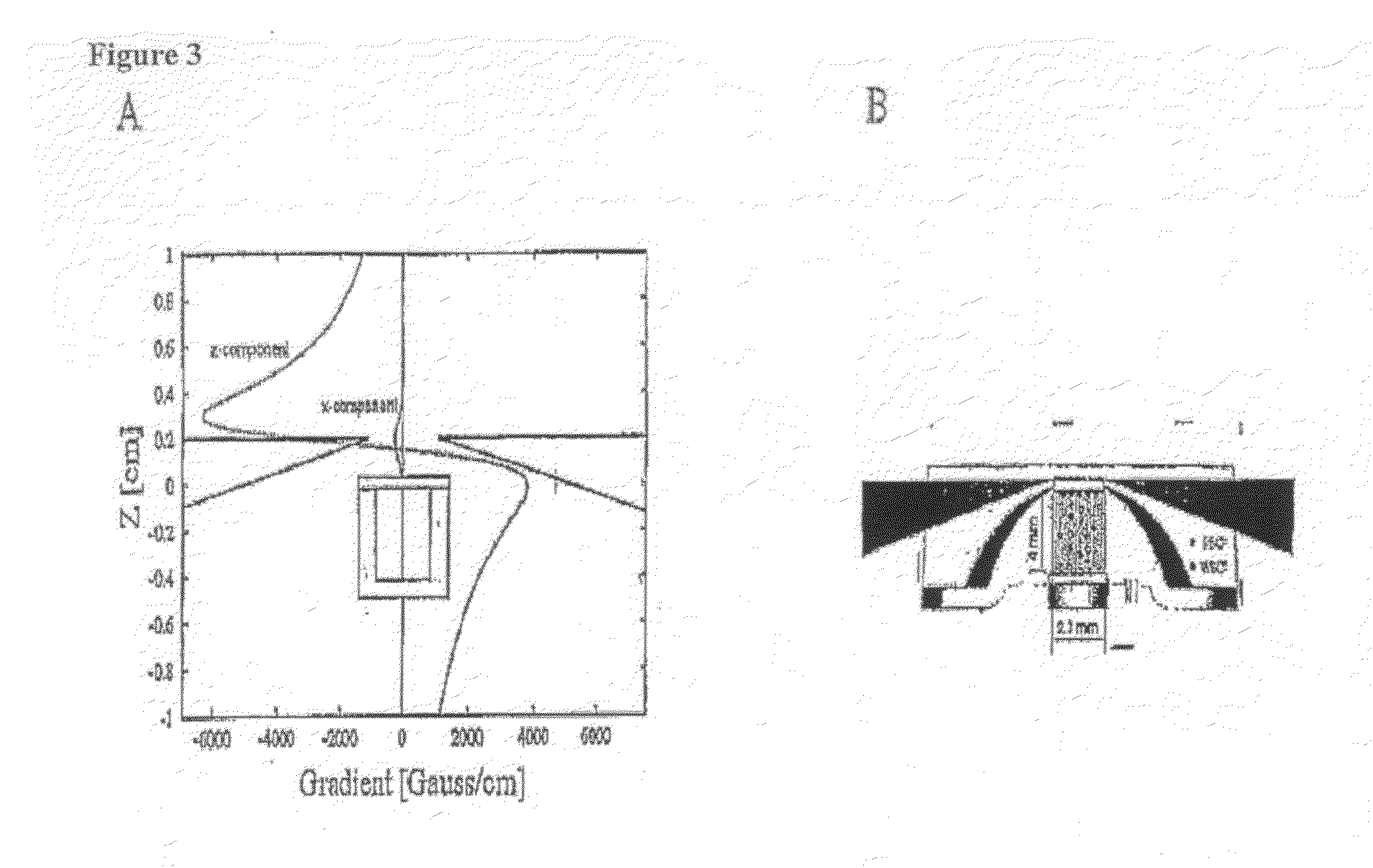
Figure 3
Abstract
Description
CROSS REFERENCE TO RELATED APPLICATIONS
[0001] This application is a non-provisional application, which is incorporated by reference herein and claims priority, in part, of U.S. Provisional Application No. 60 / 932,698, filed 16 Apr. 2007, now abandoned.FIELD OF THE INVENTION
[0002] This invention relates generally to simple and low cost electronic optical devices, methods and algorithms for enumeration of microscopic particles distributed in a two-dimensional plane. More specifically, the present invention relates to the adaptation of automated cell enumeration platforms for the direct detection of virus-infected cells in a fluid sample such as nasopharyngeal samples.BACKGROUND OF THE INVENTION
[0003] The enumeration of absolute levels of cells and their subsets in body fluids is of primary importance in determining the state of health of human beings and mammals in general. The primary analytical platform for performing such analyses is viral culture, direct immunofluorescence (DFA), or f...
Examples
example 1
Detection of Virus-Infected Cells Using Rare Cell Analysis
[0041]Objective; We studied an adapted cell detection technology, originally developed to detect ultra-rare tumor cells in circulation, to capture and detect virus-infected cells present in various cell culture systems and patient NP samples.
Relevance: Clinical diagnosis of viral infection often involves direct specimen analysis using Direct Fluorescent Antibody (DFA) followed by virus culture in susceptible cell lines. DFA is faster but is labor intensive, provides a subjective result and is not as sensitive as culture or molecular methods that may require 6 to 48 hrs for a result.
Methodology: Virus culture was done using standard methods using A549, Mink Lung, SKBr-3, and RMix (DHI) as susceptible host cell lines. Ferrofluids (FF) were prepared as colloidal suspensions of 200 nM magnetic particles coated with antibodies to specific antigen targets on cells. Suspensions of cells were fixed with cell fixative solution, washed...
example 2
Feasibility Study of a Rapid, Semi-Automated Method to Detect Respiratory Virus Infected Cells in Direct Specimens
[0042]Objective: The objective of this study was to demonstrate that a rapid, liquid specimen processing procedure could readily differentiate positive and negative cell culture in <2 min using EasyCount™, an automated fluorescent microscope with cell counting capability. EasyCount™ uses a CCD camera to visualize counter stained cells for a total cell count and interrogates those same cells to identify those that are infected with virus by detecting fluorescein.
Methods: We chose R-Mix and Influenza A (Flu) as a prototype cell culture / virus system to simulate a direct specimen scenario for automated detection purposes. Flu virus was inoculated in duplicate onto 24-well R-Mix plates, centrifuged at 700×g for 60 min, and incubated at 37° C. for 20 hr. One plate was fixed, stained and counted to confirm the viral input. The cells from the replicate plate were released and ce...