
Methods and Algorithms For Cell Enumeration in a Low-Cost Cytometer
a low-cost, cytometer technology, applied in the direction of instruments, computing, measurement devices, etc., can solve the problems of inaccessibility to routine clinical use of flow cytometry and similar complex analytical systems, lack of robustness, and exhibited rather complex results, so as to achieve low cost, low cost, and simple design
Patent Information
- Authority / Receiving Office
- US · United States
- Patent Type
- Applications(United States)
- Current Assignee / Owner
- Publication Date
- 2011-03-03
- Estimated Expiration
- Not applicable · inactive patent
Smart Images
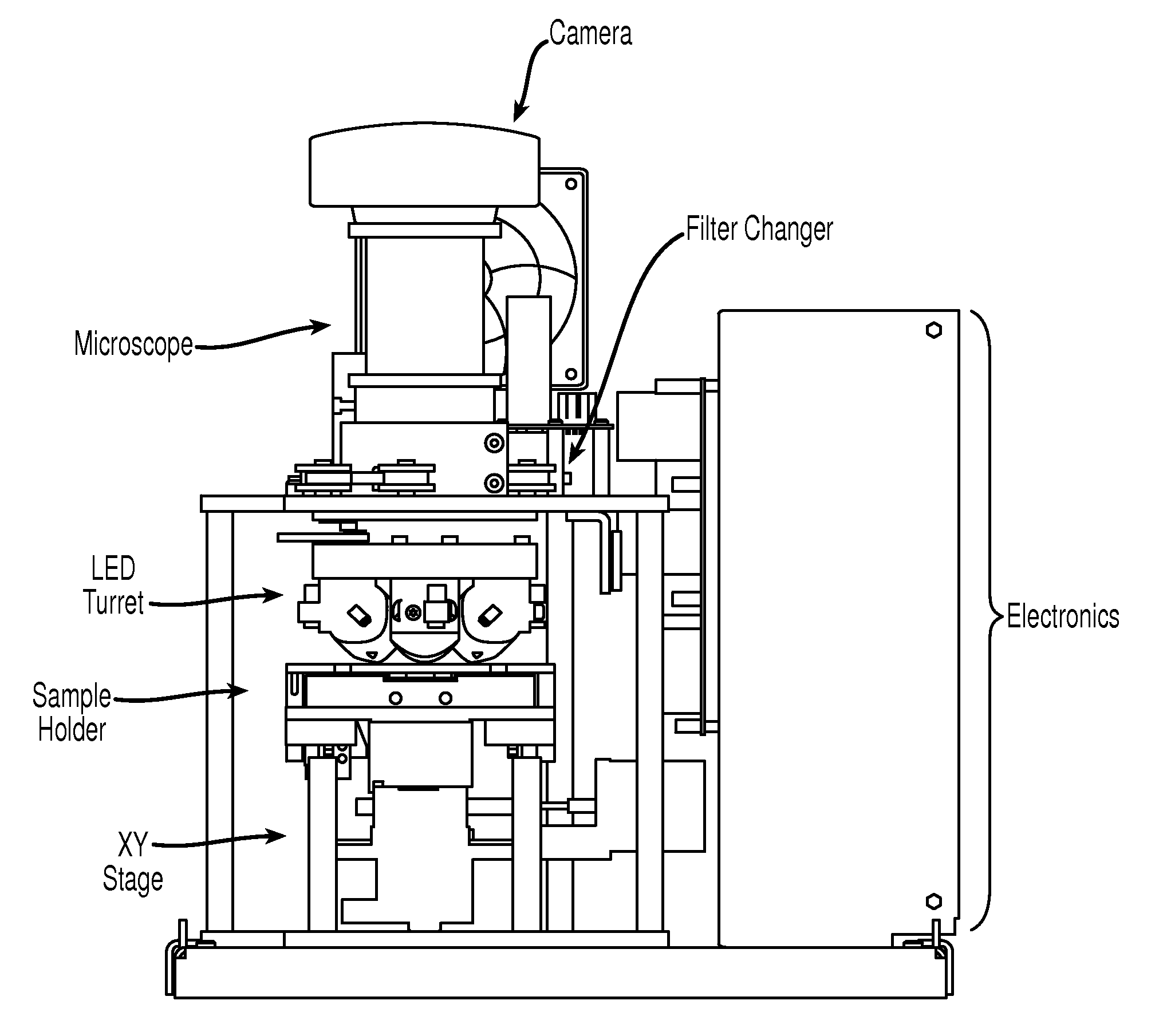
Figure 1 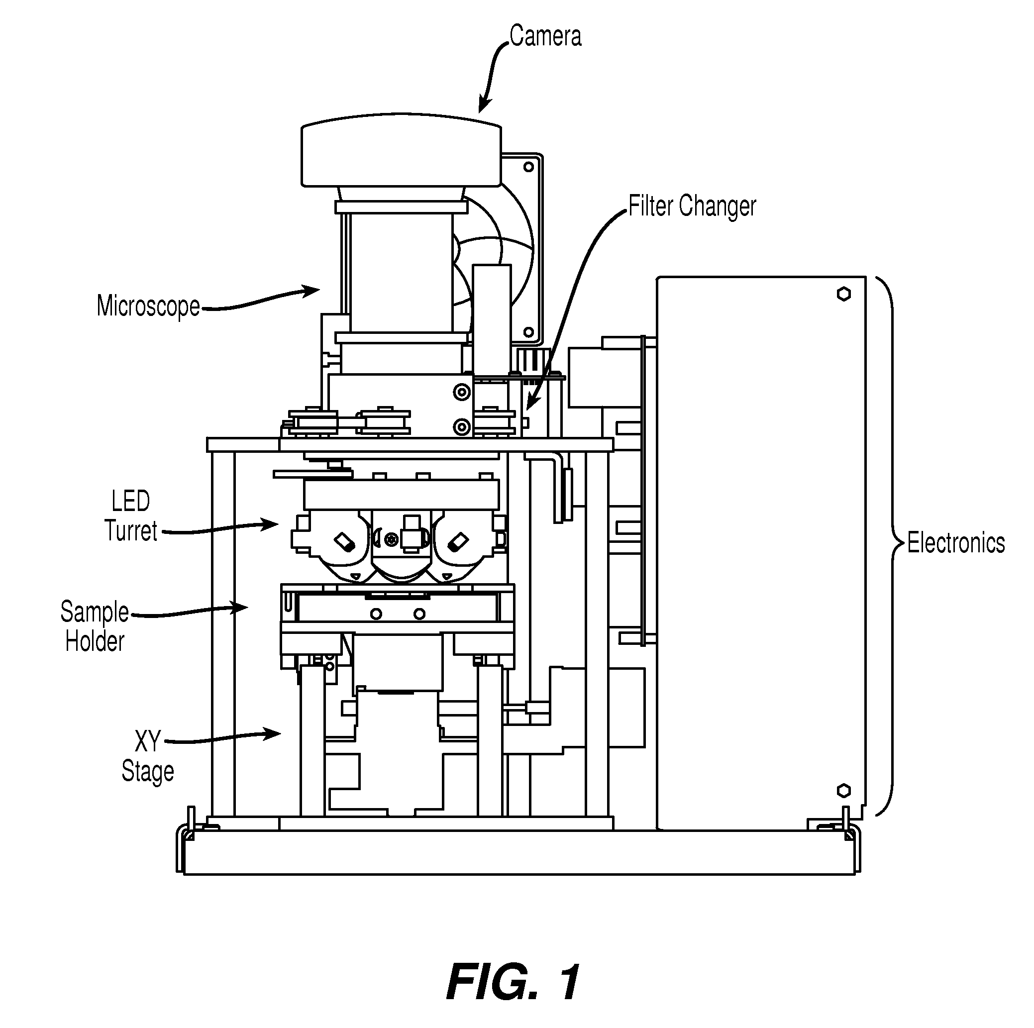
Figure 2 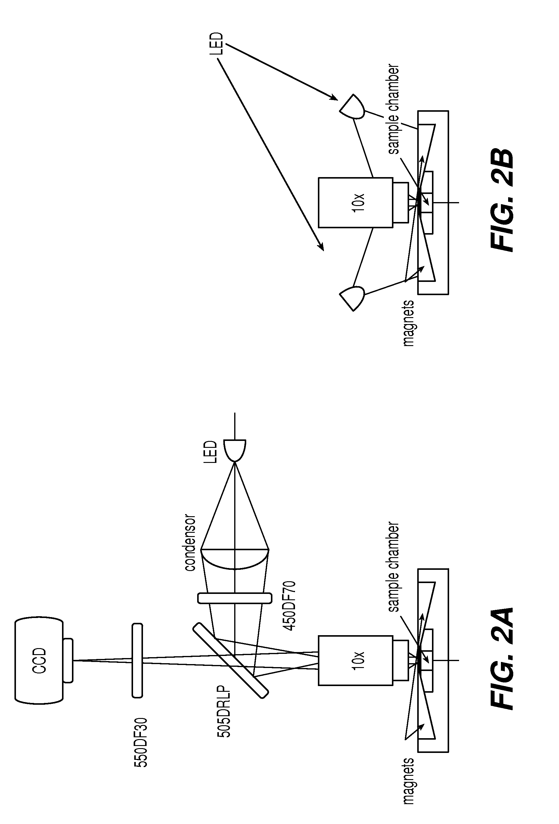
Figure 3
Abstract
Description
CROSS REFERENCE TO RELATED APPLICATIONS
[0001] This application is a divisional application from non-provisional application U.S. Ser. No. 11 / 434,321, filed 12 May 2006, which is continuation-in-part of U.S. Ser. No. 10 / 903,798, filed Jul. 30, 2004, incorporated by reference herein and claims priority, in part, of International Application PCT / US03 / 04468, filed 14 Feb. 2003, which is based upon U.S. Provisional Application No. 60 / 357,170, filed 14 Feb. 2002, now abandoned.FIELD OF THE INVENTION
[0002] This invention relates generally to simple and low cost electronic optical devices, methods and algorithms for enumeration of microscopic particles distributed in a two-dimensional plane. The novel counting techniques are particularly applicable to enumerating magnetically selected fluorescent cells in complex biological specimens such as blood by means of an inexpensive cell cytometer. Further, the present invention is applicable in the analysis of specific sample characteristics such as,...
Examples
example 1
[0048]Cell enumeration is commonly performed using expensive flow cytometry (FC) or less accurate manual count methods. Here, we present an automated microscope device to obtain absolute cell counts based on fluorescence with cell identification not prone to operator to operator variability of manual cell counts.
[0049]The device of the present invention, EASYCOUNT®, is a simple fluorescence microscope coupled to an on board microcomputer. It has a 5× objective (NA 0.1) and 4 high power LEDs for excitation at two different wavelengths. Each LED is equipped with an excitation filter and a collection lens for the stray light. The LEDs are placed next to the objective lens and illuminates the sample directly. Available illumination wavelengths are 490, 530 and 590 nm. Detection is performed by capturing a fluorescent image from a field of view of 0.95×1.3 mm on a 16 bit camera. The light is collected by the objective and selected by emission filters. The illumination and detection wavel...
example 2
[0053]Cells from the prostate cell line PC3-9 were harvested and resuspended in RPMI culture media. Cell count and viability determination by hemacytometer involved mixing cells 1:1 with trypan blue. Cells to be counted were mixed 1:1 with a cocktail containing acridine orange and ethidium bromide. Samples were then loaded onto a slide, placed on the EASYCOUNT® platform, and viable and non-viable counts were obtained. Cells at concentrations ranging from 2×10⁴ / ml through 5×10⁶ / ml were analyzed for total cell counts using both the hemacytometer and EASYCOUNT®. Regression analysis of the data showed an R2 of 0.999 with a slope of 1.2. At the upper end of the titration curve, hemacytometer counts required an additional dilution step which was not necessary with EASYCOUNT®. Regression analysis showed an R2 of 0.97 with a slope of 0.99. Correlation of sample viability involved making mixtures of cells with viabilities of approximately 0, 25, 50, 75, and 100%. Non-viable cells were prepar...
example 3
[0054]Counting residual leukocytes in leukodepleted blood products is done using either a specialized hemacytometer (NAGEOTTE™) or by flow cytometry on nucleic acid-stained samples. NAGEOTTE™ counting is tedious and labor-intensive. Flow cytometry requires expensive instrumentation, a skilled operator, and has low sample throughput. The fluorescent imaging system of the present invention is useful for counting low numbers of residual leukocytes in leukodepleted blood products.
[0055]One hundred microliters of blood were mixed with anti-CD45-ferrofluid and a fluorescent stain to visualize the cells. A variety of fluorescents stains were evaluated among which nucleated acid dyes and anti-CD45-PE. Leukocytes were magnetically mounted in a cartridge, used in the fluorescent imaging device CELLTRACKS®, and then the cartridge was placed in a device for magnetic orientation of magnetized targets, MAGNEST®, and imaged using either an EASYCOUNT® cell counting device or a fluorescent microscop...