
Calibration method for radiation detector, and particle therapy system
Patent Information
- Authority / Receiving Office
- US · United States
- Current Assignee / Owner
- Publication Date
- 2013-08-29
Smart Images
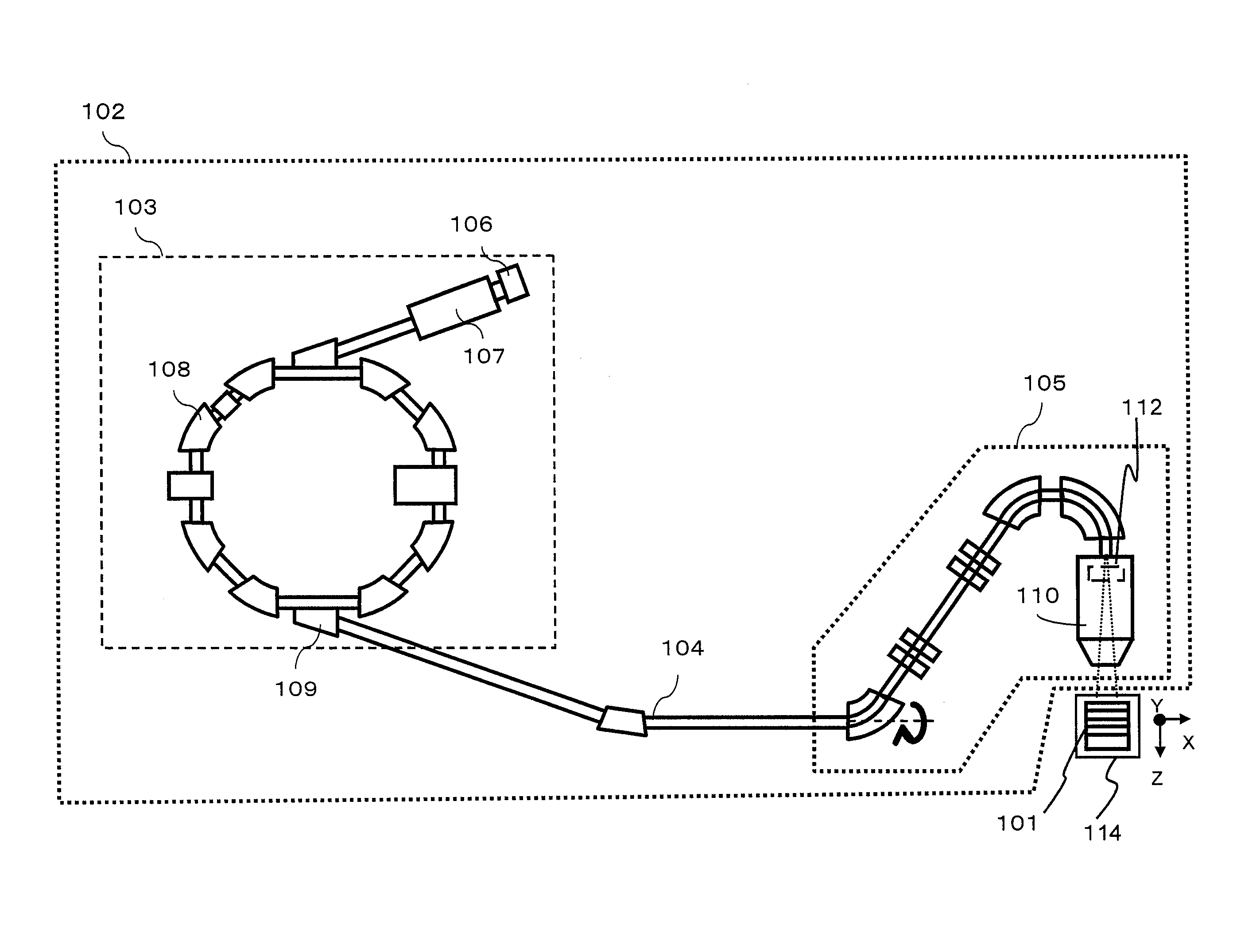
Figure 1 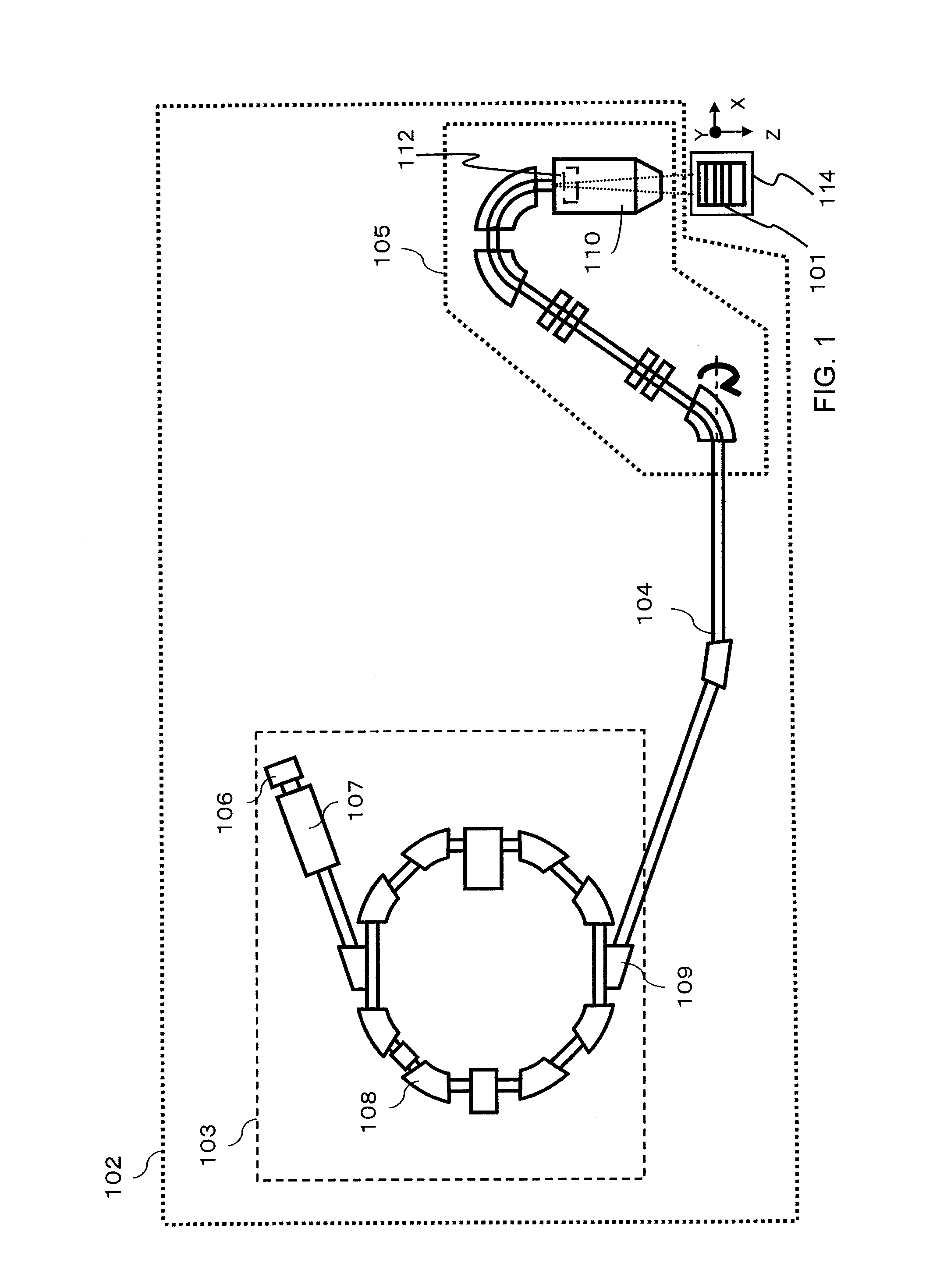
Figure 2 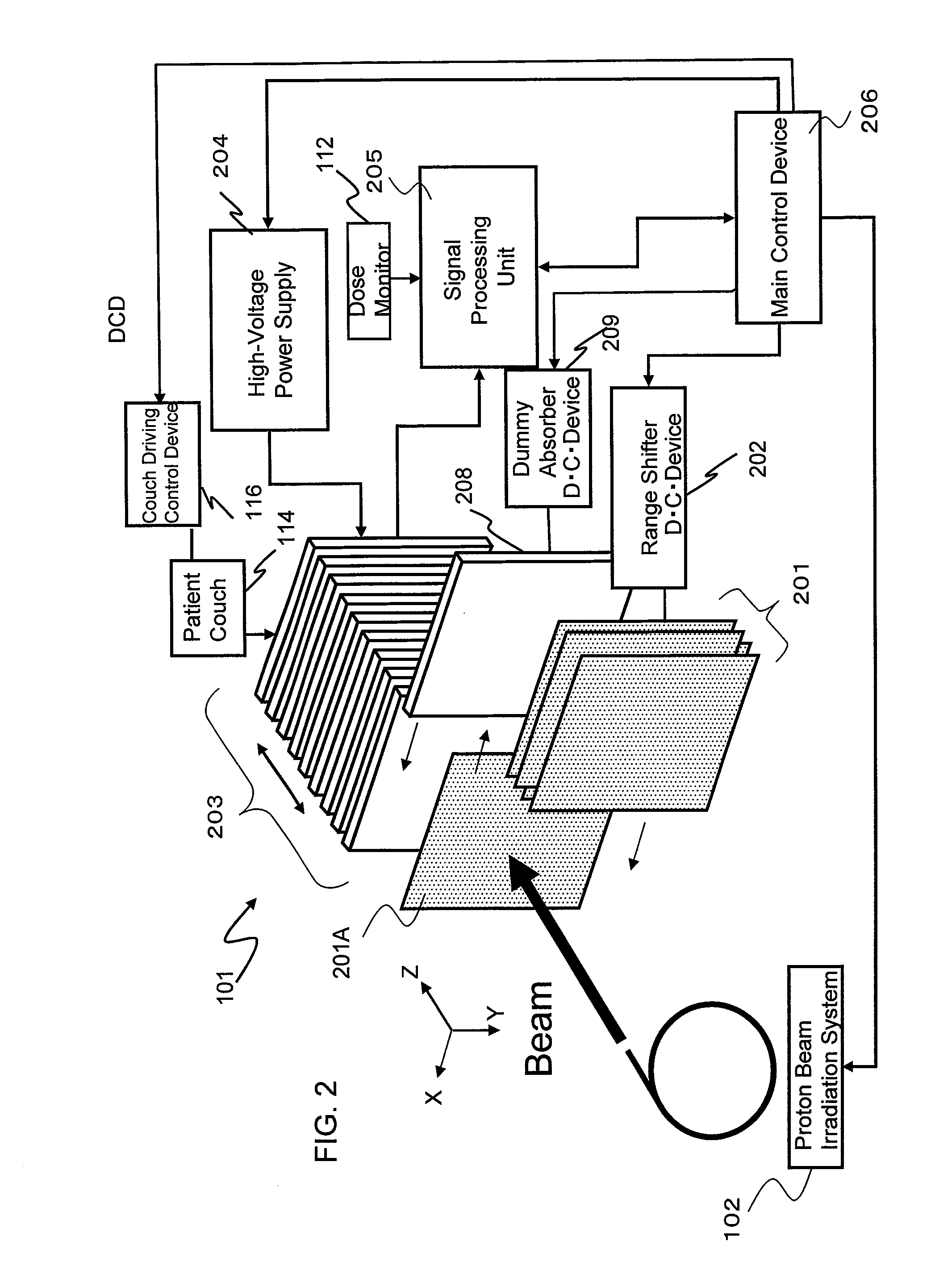
Figure 3
Abstract
Description
BACKGROUND OF THE INVENTION
[0001] 1. Field of the Invention
[0002] The present invention relates to a method for calibrating a radiation detector, and to a particle therapy system.
[0003] 2. Description of the Related Art
[0004] A scanning method is becoming widespread in particle therapy. The scanning method is a technique used to divide a target region into very small segments, called spots, and irradiate each spot independently with a narrow beam having a small diameter of 1σ=3 to 20 mm. The irradiation is stopped once a predetermined dose has been delivered to the spot, and after that, a next spot is scanned with the same beam. Scanning magnets are used when the beam is to be scanned in a direction perpendicular to a direction in which the beam travels (hereinafter, this scanning direction is referred to as the lateral direction, and likewise the traveling direction of the beam, as the depth direction). After the predetermined dose has been delivered to a certain depth in all spots, th...
Examples
Embodiment Construction
[0019]Particle therapy systems that employ the scanning method are adjusted to within scan position errors of ±1 mm so that the system scans a beam laterally across an indicated position. These systems are also adjusted to within beam range errors of ±1 mm to ensure that the beam reaches an indicated depth level. The range denotes a maximum depth that the beam can reach when it travels through the medium, and this value depends upon incident energy of the beam.
[0020]To confirm adjustment results on the particle therapy system, a radiation detector measures the scanning position and Bragg curve of the beam. The Bragg curve is a distribution of linear energy transfer (LET) in the depth direction, expressed in terms of [J / m], and depends upon the incident energy of the beam. More specifically, LET is the energy that the particle beam passing through the medium transfers thereto while traveling through a unit distance in the depth direction. The beam's reachable depth is calculated from...