
Inhibition of aminocylase 3 (AA3) in the treatment of cancer
Patent Information
- Authority / Receiving Office
- US · United States
- Current Assignee / Owner
- Publication Date
- 2020-07-02
Smart Images
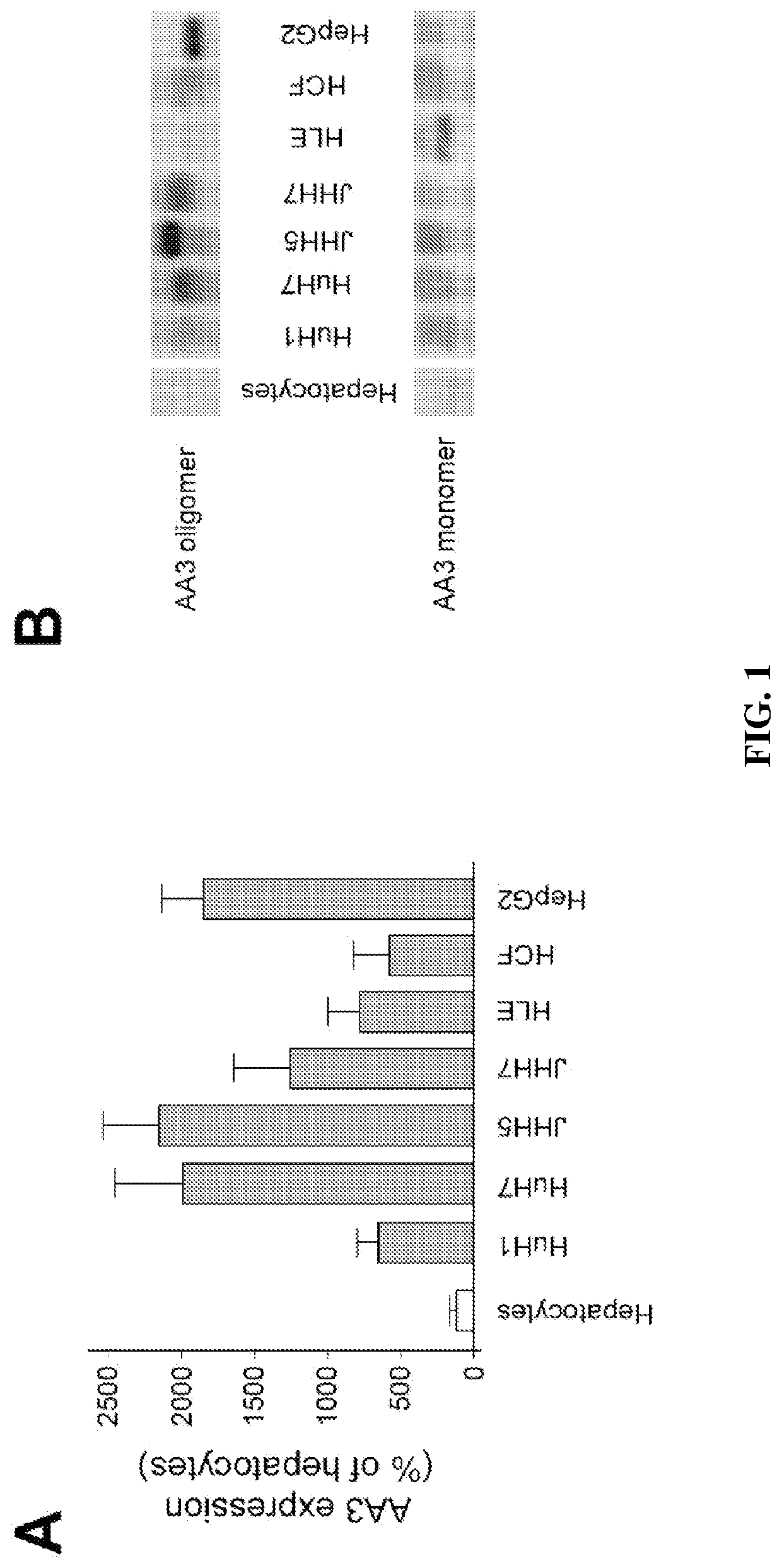
Figure 1 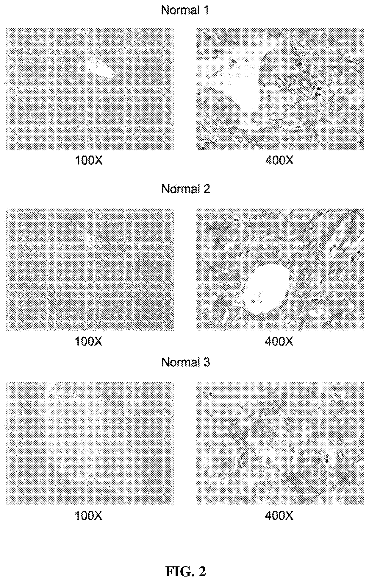
Figure 2 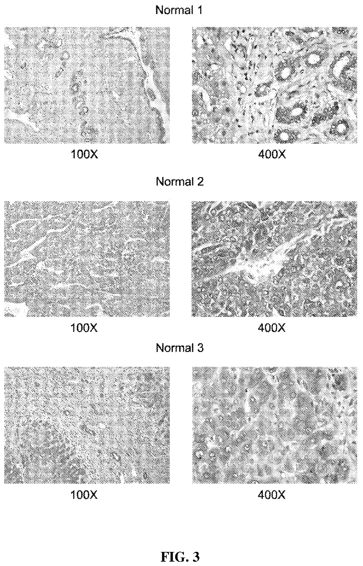
Figure 3
Abstract
Description
CROSS-REFERENCE TO RELATED APPLICATIONS
[0001] This application claims the benefit of priority of U.S. Provisional Patent Application No. 62 / 559,170 filed Sep. 15, 2017, which is hereby incorporated by reference in its entirety.BACKGROUND OF THE INVENTION1. Field of the Invention
[0002] The present disclosure relates generally to the fields of biology and medicine. Particularly, it concerns chemical compounds, methods, and compositions involving those compounds. More particularly the present invention relates to cancer, including hepatocellular carcinoma (HCC), pancreatic and colon cancer. The present invention also relates to methods and compositions of treating patients, including patients with HCC, pancreatic and colon cancers.2. Background Art
[0003] Ras proteins are small GTPases and key regulators of diverse signal transduction pathways controlling cell growth, differentiation and apoptosis. They are also common oncogenes mutated in ˜20% of all cancers [1-16]. Upregulation of Ras vi...
Examples
example 1
n of AA3 in Primary Hepatocytes, HCC Cell Lines, Normal and HCC Livers
[0172]All studied HCC cell lines (HuH1, HuH7, JHH5, JHH7, HLE, HCF and HepG2) expressed AA3 (FIG. 1). AA3 expression in HCC cell lines was 5 to 20 times greater than in normal primary hepatocytes (FIG. 1A). There were two AA3 bands of ˜35 and 90-100 kDa (FIG. 1B). The immunostaining was blocked by pre-incubation of HR-C1 antibody with purified human AA3. The higher mobility band corresponded to the AA3 monomer (˜35 kDa) that has been reported to be expressed in normal mammalian kidney and liver [32, 35-38]. The ˜90-100 kDa band was detected in most HCC cell lines but not in normal hepatocytes (FIG. 1).
[0173]The inventors studied next the protein expression level of AA3 in normal liver and HCC livers. As shown in FIG. 2, AA3 protein is expressed mostly in hepatocytes with weak expression in biliary epithelial cells (FIG. 2). AA3 expression in normal liver was negative (−) to minimal (+) and was moderate (++) to hig...
example 2
tylates NAFC and NAGGC
[0174]Human AA3 efficiently deacetylates NAFC (FIG. 4a-d) and NAGGC (FIG. 4e-h) generating FC and GGC respectively. Kinetic parameters for these substrates (Km: 0.025 and 0.14 mM; kcat: 2 and 7.2 s−1 for NAFC and NAGGC, respectively) are similar to the other AA3 substrates, N-acetylated aromatic amino acids and mercapturic acids [30-33, 38,39].
[0175]AA1 (EC 3.5.1.14), in addition to AA3, has been reported to deacetylate certain mercapturic acids [36], although the substrate specificity of AA1 differed from AA3 [30-33, 39]. Therefore, we determined whether AA1 deacetylates NAFC and NAGGC. In experiments conducted by the inventors, rat AA1 that has high sequence identity with human AA1 did not significantly deacetylate these mercapturates: the specific activity was less than 0.1% of the corresponding value of AA3 and therefore the exact activity value could not be estimated (FIG. 5a).
[0176]Since HCC cell lines express the ˜90-100 kDa form that may change the abil...
example 3
AA3 Inhibitors on FC and NAFC Levels in HCC Cell Lines
[0178]The inventors' data suggest that AA3 deacetylates and therefore decreases the level of NAFC and NAGGC in HCC cells, and conversely AA3 inhibition should increase these levels. Accordingly, we measured NAFC and FC levels in HCC cells before and after treatment with AA3 inhibitors [33]. In untreated HepG2 and HuH7 cell lines, both FC and NAFC were detected; the level of FC in both cell lines was significantly higher than NAFC (FIG. 6). The FC level in HepG2 cells was higher than in HuH7 cells whereas the NAFC level in both cell lines was similar. Correspondingly the [FC] / [NAFC] ratio in HepG2 cells was slightly smaller (˜2.6) then HepG2 cells (˜3.7). Treatment with AA3 inhibitors 10 and 11 significantly increased the level of NAFC in both cell lines; the FC level was not significantly changed in HepG2 cell line, and in HuH7 cell lines decreased by approximately 50%. In the HepG2 cell line, the [FC] / [NAFC] ratio decreased from...