
Co-delivery of stimulatory and inhibitory factors to create temporally stable and spatially restricted zones
a technology of stimulatory factors and inhibitory factors, applied in the field of drug delivery, can solve the problems of difficult spatial cue cellular discrimination, achieve the effects of preventing the negative effects of initial bursts, promoting cell-based maintenance of tissue structure and function, and halting or retarding disease progression or age-related tissue changes
Patent Information
- Authority / Receiving Office
- US · United States
- Patent Type
- Patents(United States)
- Current Assignee / Owner
- Publication Date
- 2017-07-04
Smart Images
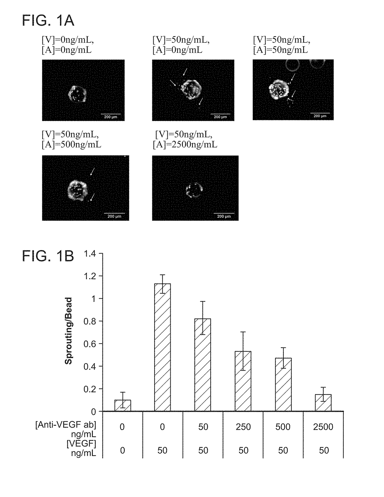
Figure 1 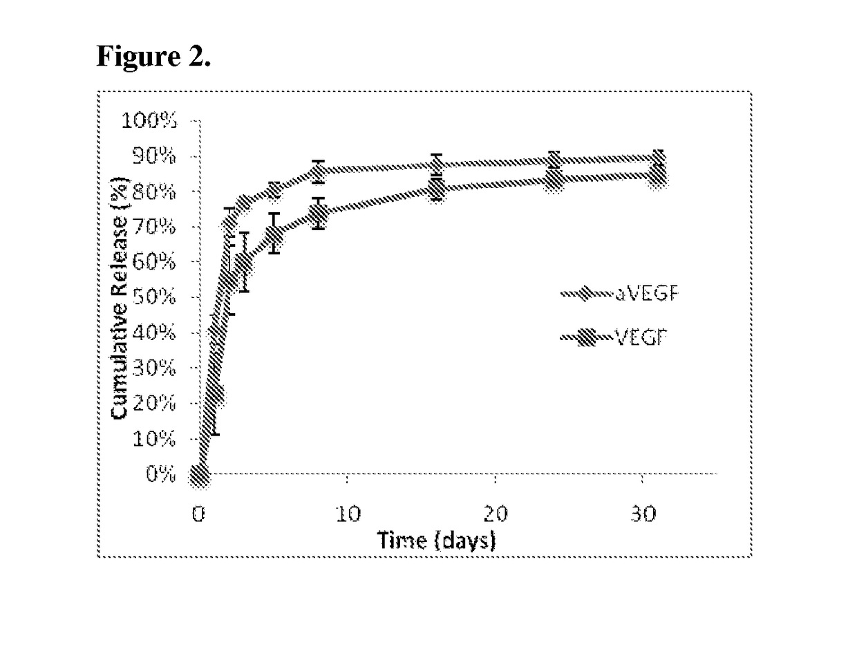
Figure 2 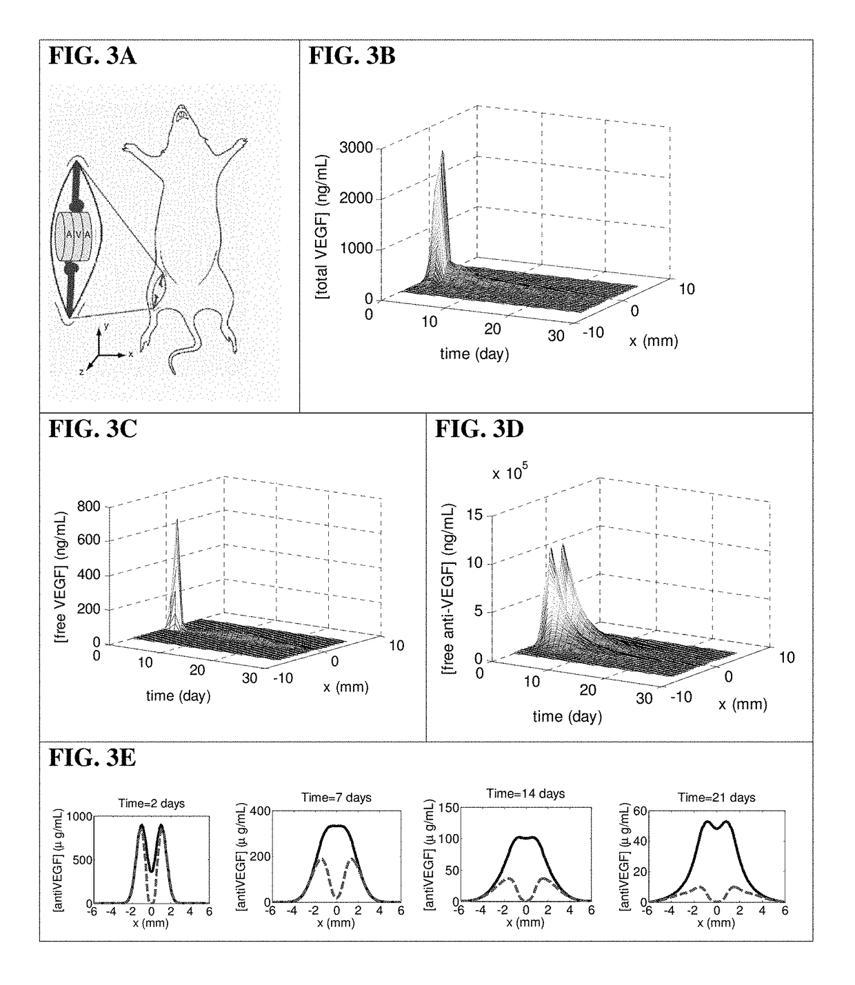
Figure 3
Abstract
Description
RELATED APPLICATIONS
[0001] This application is a national stage application, filed under 35 U.S.C. §371, of International Application No. PCT / US2011 / 042051, filed Jun. 27, 2011, which claims the benefit of U.S. Provisional Patent Application No. 61 / 358,499, filed on Jun. 25, 2010, the contents of which are incorporated herein by reference in their entireties.STATEMENT AS TO FEDERALLY SPONSORED RESEARCH
[0002] This invention was made with Government support under National Institutes of Health awards R01HL069957 and R37DE013033. The Government has certain rights in the inventionFIELD OF THE INVENTION
[0003] This invention relates generally to the field of drug delivery.BACKGROUND OF THE INVENTION
[0004] Nature frequently utilizes opposing factors to create a stable activator gradient to robustly control pattern formation. Specifically, during developmental processes, tight spatial regulation often results from the combined action of stimulatory and inhibitory factors. In such reaction scheme...
Examples
example 1
VEGF and Anti-VEGF on Angiogenesis
[0119]The relation between VEGF and anti-VEGF concentrations on angiogenesis was first evaluated using a common in vitro sprouting assay, in order to quantitatively determine the appropriate doses of the two factors for subsequent in vivo studies.
Cell Culture and In Vitro Sprouting Assay
[0120]Briefly, dermal human vascular endothelial cells (HMVECs) were purchased from Lonza (CC-2543) and cultured to confluence at 37° C. and 5% CO2 in microvascular endothelial cell growth medium-2 (EGM-2MV) (Lonza) containing all supplements. Angiogenic activity of endothelial cells was assessed using a modification of a widely used in vitro sprouting assay (Nehls, V., and Drenckhahn, D. 1995 Microvasc Res 50, 311-322). Briefly, dextran beads microcarriers (Cytodex 3) with a dry weight of 50 mg were swollen in PBS and autoclaved. The microcarriers were washed in EGM-2MV medium and seeded with 3×10⁶ HMVECs in a spinner flask. The microcarriers were stirred for 2 minu...
example 2
cal Model of Protein Distribution
[0126]In order to design appropriate encapsulated doses of VEGF and anti-VEGF to create spatially defined angiogenic regions, mass transport PDEs of the proteins in the scaffolds and the underlying tissues were simulated.
[0127]Briefly, a computational model was generated to depict the concentration profiles of free VEGF anti-VEGF, and VEGF complexed with anti-VEGF. This model accounted for diffusion, release from scaffolds, binding kinetics, and protein degradation. The governing equations of the VEGF and anti-VEGF concentrations inside the scaffold and underlying muscle were:
[0128]∂c1∂t=D1∇2c1-k1c1+f1-konc1c2+koffc3∂c2∂t=D2∇2c2-k2c2+f2-konc1c2+koffc3∂c3∂t=D3∇2c3+konc1c2-koffc3whereci=concentrationci(x,y,z,t=0)=0;∀i,x,y,zfi={releasefunction,insidescaffold0,insidemusclei={1freeVEGF2freeantiVEGF3VEGF-antiVEGFcomplexD1=7×10⁻⁷cm2s=EffectiveinterstitialdiffusioncoefficientofVEGF165D2=3.2× 10⁻⁹cm2s= Effe...
example 3
Regulated Angiogenesis In Vivo
[0135]To test the ability of this system to provide spatial control over angiogenesis, scaffolds were subsequently implanted into the ischemic hindlimbs of SCID mice.
Mouse Model of Hindlimb Ischemia
[0136]Scaffolds were implanted in 6-week-old SCID mice (Taconic, Hudson, N.Y.) that had undergone unilateral ligation of hindlimb blood vessels to create a severe model of hindlimb ischemia (Sun et al., 2005 Pharm Res 22, 1110-1116). The SCID model was chosen because it offered a stable loss of perfusion over weeks and the angiogenic effects from inflammation were reduced. Briefly, animals were anesthetized by IP injection of a 7:1 mixture of ketamine and xylazine. The targeted hindlimb was shaved and sterilized with ethanol prior to making an incision through the dermis. Ligation sites were made on the external iliac artery and vein, and on the femoral artery and vein using 5-0 Ethilon (Ethicon, Somerville, N.J.). The vessels were severed between the ligatio...