Space compound imaging method in ultrasonic diagnosis

A technology of spatial compounding and imaging methods, which is applied in the directions of acoustic wave diagnosis, infrasonic wave diagnosis, ultrasonic/sonic wave/infrasonic wave diagnosis, etc. It can solve the problems of reducing the contrast resolution of ultrasonic image D and the sense of hierarchy of tissue structure, so as to achieve a good sense of hierarchy, Strengthen the beam signal strength and enhance the effect of information

Inactive Publication Date: 2011-05-04
VINNO TECH (SUZHOU) CO LTD
View PDF3 Cites 11 Cited by
  • Summary
  • Abstract
  • Description
  • Claims
  • Application Information

AI Technical Summary

Problems solved by technology

[0002] In the ultrasonic diagnostic instrument, the existing spatial composite imaging method includes: performing multiple ultrasonic scans in different directions on the target tissue, in the figure 1 In this method, the ultrasonic image A is obtained by sweeping to the left, the ultrasonic image B is obtained by the forward sweep, and the ultrasonic image C is obtained by sweeping to the right, and then the amplitude of the three image points with the same coordinates is linearly Averaging, the ultrasonic image D composed of new image points is obtained. The texture structure of the ultrasonic image D is fine, the spatial organization structure is rich, and the speckle noise is less. However, this linear average is performed on images scanned in different time frames and directions in the same space. The method will reduce the contrast resolution of the ultrasound image D and the layering of the tissue structure, so that there is not much useful information related to the diagnostic site in the ultrasound image D

Method used


Image

  • Space compound imaging method in ultrasonic diagnosis
  • Space compound imaging method in ultrasonic diagnosis
  • Space compound imaging method in ultrasonic diagnosis

Examples


Embodiment Construction

[0012] The structure of the present invention will be further described below in conjunction with the accompanying drawings.

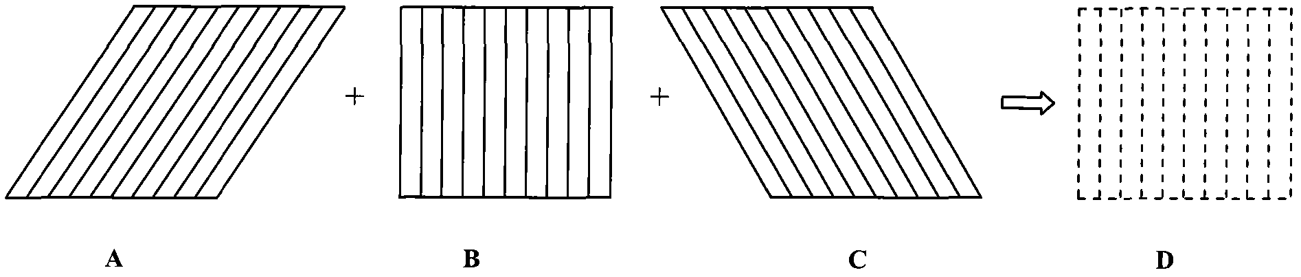
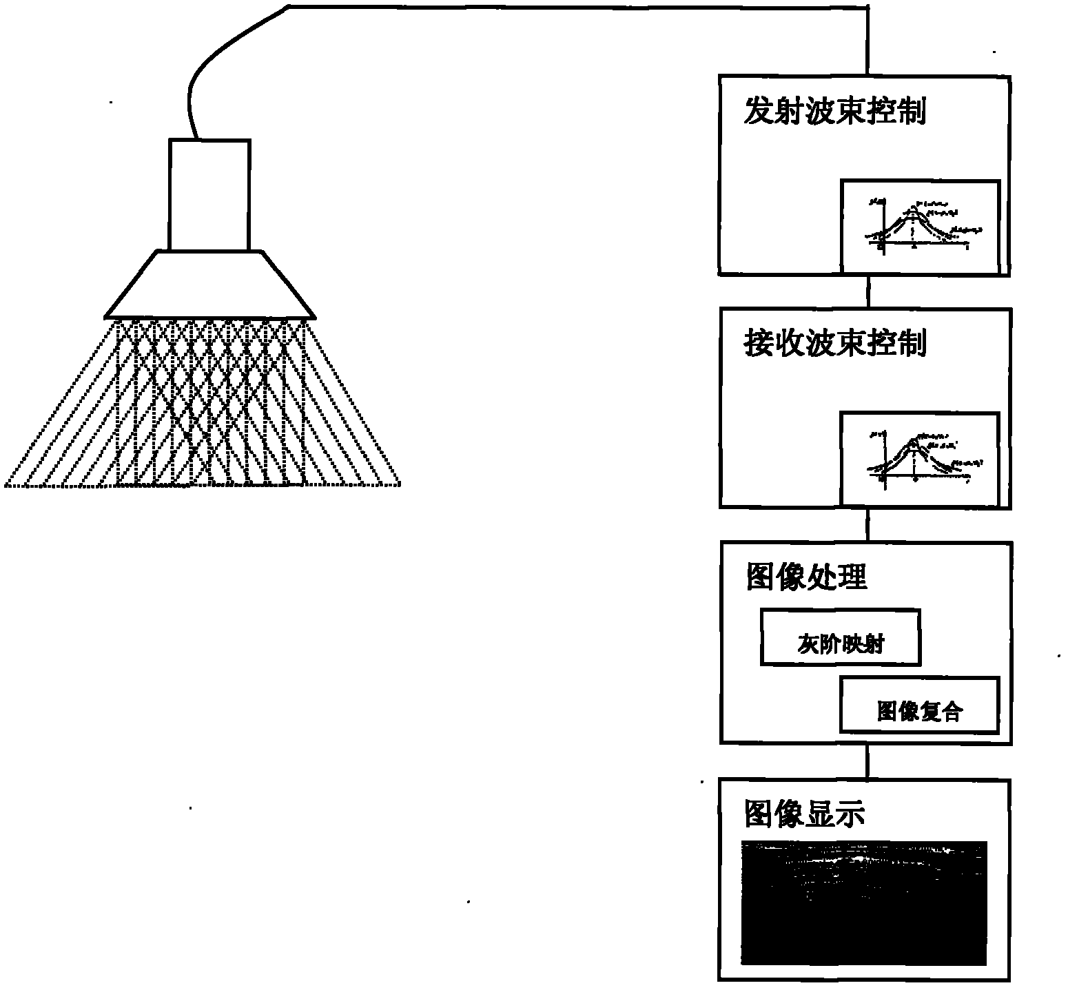
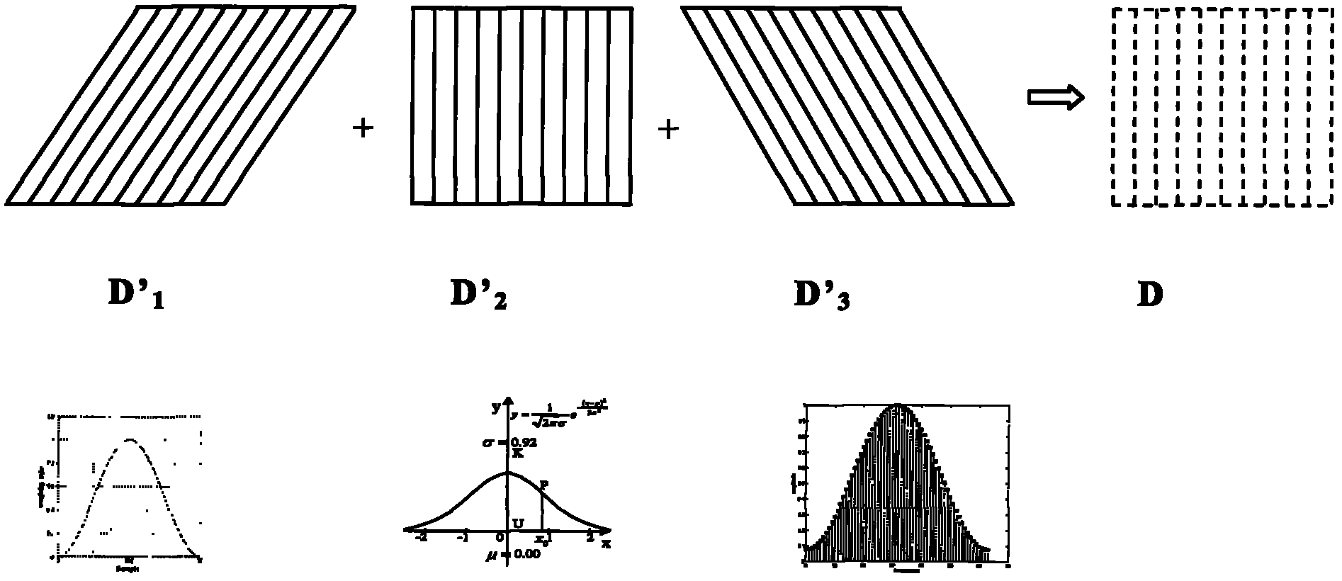
[0013] exist Figure 2-3 Among them, the spatial compound imaging method of ultrasonic diagnosis, through the spatial compound superposition of ultrasonic images scanned in multiple different directions, obtains high-quality ultrasonic images. When scanning the target tissue in different directions, due to the different scanning angles and the slight difference in scanning time, the actual ultrasonic echo signals obtained in different scanning directions are not the same, which requires us to analyze different angles in the same space. In order to obtain the best spatial composite imaging, different signal and image processing are performed on the image frames at different times and at different times. Specifically, the apodization window control is performed on the beam signals of the transmitting and receiving channels in N (N>=2) different directio...


PUM


Abstract

The invention discloses a space compound imaging method in ultrasonic diagnosis. The method has the following advantages: window function processing is carried out on wave beam signals in transmitting and receiving channels during ultrasonic scanning in N directions respectively, then the obtained initial ultrasonic image data in N directions are processed through a histogram equalization algorithm or matching algorithm, and then space compound imaging is carried out on the processed ultrasonic image data to obtain the final ultrasonic image; the technicians in the field need to carry out debugging; and appropriate window function processing is selected, thus enhancing the wave beam signal intensity of the diagnosed part in the target tissue, and an appropriate histogram equalization algorithm or matching algorithm is conductive to further elevating the image data on the diagnosed part and enhancing the information of the diagnosed part in the final ultrasonic image, so the contrast resolution of the final ultrasonic image and hierarchy of the tissue structure are better and more useful information of the diagnosed part is displayed.

Description

technical field [0001] The invention relates to a spatial compound imaging method for ultrasonic diagnosis. Background technique [0002] In the ultrasonic diagnostic instrument, the existing spatial composite imaging method includes: performing multiple ultrasonic scans in different directions on the target tissue, in the figure 1 In this method, the ultrasonic image A is obtained by sweeping to the left, the ultrasonic image B is obtained by the forward sweep, and the ultrasonic image C is obtained by sweeping to the right, and then the amplitude of the three image points with the same coordinates is linearly Averaging, the ultrasonic image D composed of new image points is obtained. The texture structure of the ultrasonic image D is fine, the spatial organization structure is rich, and the speckle noise is less. However, this linear average is performed on images scanned in different time frames and directions in the same space. This method will reduce the contrast resol...

Claims


Application Information

Patent Timeline
Patent Type & Authority Applications(China)
IPC IPC(8): A61B8/00G06T5/50
Inventor 田林
Owner VINNO TECH (SUZHOU) CO LTD
Why Eureka
Try Eureka