Method and system for dividing up three-dimensional medical image of abdominal organ

A medical image and abdominal technology, applied in the field of 3D medical image segmentation method and system of abdominal organs, can solve problems such as blurred boundaries of medical image segmentation, and achieve the effect of benefiting medical analysis and diagnosis, increasing calculation time, and improving quality

Inactive Publication Date: 2012-01-04
SHENZHEN YORATAL DMIT
View PDF3 Cites 17 Cited by
  • Summary
  • Abstract
  • Description
  • Claims
  • Application Information

AI Technical Summary

Problems solved by technology

[0006] In view of the above-mentioned defects, the object of the present invention is to provide a method and system for segmenting three-dimensional medical images of abdominal organs. Aiming at the characteristics of blurred boundaries of abdominal organs, users can interactively input blurred boundaries to solve the problem of blurred boundaries in medical image segmentation. The problem

Method used


Image

  • Method and system for dividing up three-dimensional medical image of abdominal organ
  • Method and system for dividing up three-dimensional medical image of abdominal organ
  • Method and system for dividing up three-dimensional medical image of abdominal organ

Examples


Embodiment Construction

[0043] In order to make the object, technical solution and advantages of the present invention clearer, the present invention will be further described in detail below in conjunction with the accompanying drawings and embodiments. It should be understood that the specific embodiments described here are only used to explain the present invention, not to limit the present invention.

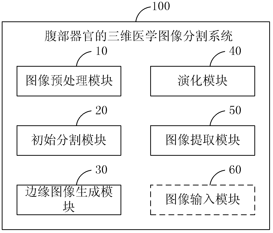
[0044] figure 1 It is a structural schematic diagram of the three-dimensional medical image segmentation system of abdominal organs in the present invention. The three-dimensional medical image segmentation system 100 mainly includes an image preprocessing module 10, an initial segmentation module 20, an edge image generation module 30, an evolution module 40 and an image extraction module 50, of which:

[0045]The image preprocessing module 10 is used for preprocessing the medical sequence images of abdominal organs. The main purpose of the preprocessing is to smooth the images and remove noise i...


PUM


Abstract

The invention discloses a method and a system for dividing up a three-dimensional medical image of an abdominal organ. The method comprises the following steps of: preprocessing a medical sequence image of the abdominal organ; selecting a seed point from the preprocessed medical sequence image, and using a pixel point in a preset neighborhood range of the seed point as an initial division region of the abdominal organ; acquiring an edge calculation image of the preprocessed medical sequence image according to an edge stop function, and receiving a feebleness edge drawing instruction of a user to obtain an edge image of the abdominal organ by combining the feebleness edge drawing instruction with the edge calculation image; performing level set function evolution to update a velocity function by combining a level set velocity function with the edge image; and extracting the velocity function to obtain a target region. Therefore, according to the invention, the problem of fuzzy boundary in medical image division is solved by using a user interaction type input boundary fuzzy boundary, and the quality of the three-dimensional division image of the abdominal organ is improved.

Description

technical field [0001] The invention relates to the field of medical image segmentation, in particular to a three-dimensional medical image segmentation method and system for abdominal organs. Background technique [0002] Medical image segmentation refers to the segmentation of the region of interest or lesion in the medical image. Its purpose is to efficiently determine the clinical morphological features that are crucial to the patient, so as to facilitate the observation and analysis of the morphological features and pathological condition. Organ segmentation is a fundamental component of computer-aided surgical planning and treatment, such as tumor resection, organ transplantation, etc. are based on organ segmentation, and can also be used for diagnostic and detection purposes. Most abdominal organ segmentation methods are basically based on CT (Computed Tomography, computerized tomography) sequence images to perform image segmentation in two-dimensional or three-dimen...

Claims


Application Information

Patent Timeline
Patent Type & Authority Applications(China)
IPC IPC(8): G06T5/00
Inventor 张磊黄健施万利
Owner SHENZHEN YORATAL DMIT
Why Eureka
Try Eureka