3D imaging and fusion method based on pathological slice scanning device

A technology of pathological slices and scanning devices, which is applied in the field of biomedicine and can solve the problems of not being able to meet the details of scanned images and not having 3D

Inactive Publication Date: 2015-12-02
曲阜裕隆生物科技有限公司
View PDF6 Cites 13 Cited by
  • Summary
  • Abstract
  • Description
  • Claims
  • Application Information

AI Technical Summary

Problems solved by technology

[0003] However, the existing pathological slice scanning devices do not have 3D effects, which makes it impossible to adjust the focal length up a...

Method used


Image

  • 3D imaging and fusion method based on pathological slice scanning device
  • 3D imaging and fusion method based on pathological slice scanning device
  • 3D imaging and fusion method based on pathological slice scanning device

Examples


Embodiment Construction

[0038] Below in conjunction with accompanying drawing, the present invention is described in further detail:

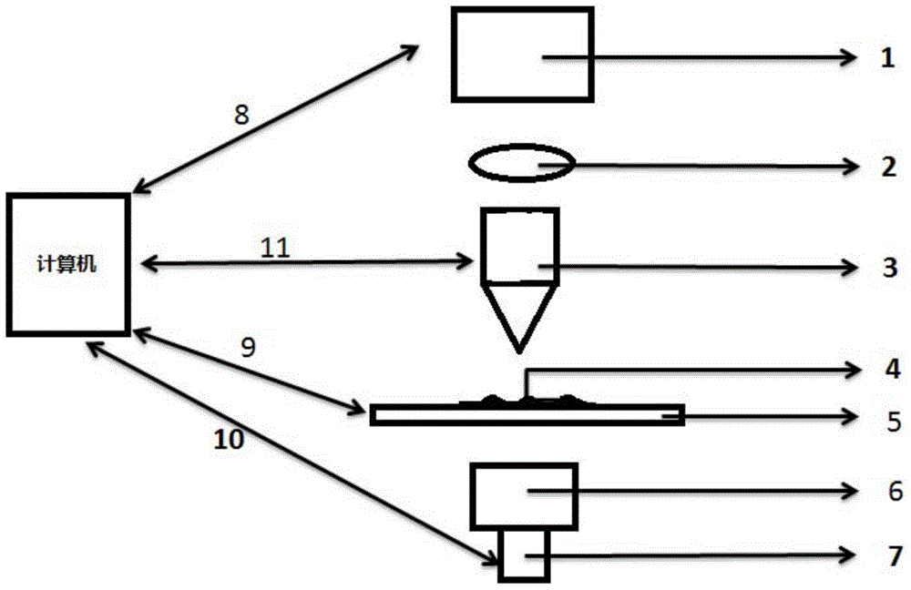
[0039] Pathological slide scanning devices such as figure 1 As shown, it includes a camera device for collecting pathological slice images, that is, a camera 1; an imaging mirror 2 for optical path imaging; an objective lens 3 for enlarging images, which can move up and down for camera focusing; pathological slice 4, that is, scanning Object; scanning platform 5, used to move pathological slices to move in X, Y directions; condenser lens 6, gather light together; light source 7: provide light source as LED light, figure 1 8-11 in the figure means that the PC communicates with the four modules, the PC controls the four modules and the four modules return data to the PC.

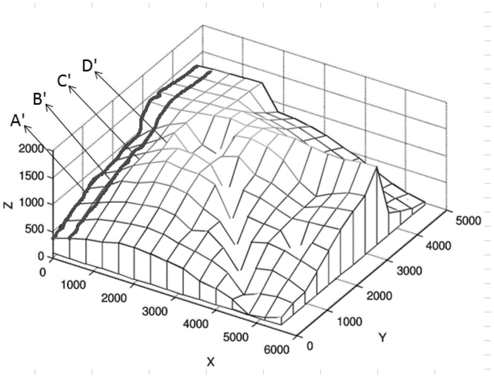
[0040] figure 2 Shown is the movement trajectory diagram of the platform XYZ axis. Calculate the scanning curve by measuring the focal point. The trajectory represented by A' is the motion curve ...


PUM


Abstract

The invention provides a 3D imaging and fusion method based on a pathological slice scanning device and belongs to the biomedicine field. Through the pathological slice scanning device, a 3D image of each focal plane is simulated. Through the fusion method, the 3D images on the different focal planes are fused on one image. Finally, data of each focal plane layer and data of a fusion layer are combined into one 3D image. By using the method of the invention, a problem that the scanned image losses details of the image itself during a scanning process of the pathological slice scanning device is solved; a 3D effect can be conveniently and concisely watched without a microscope and the above situation is simulated effectively through the pathological slice scanning device; and through a fusion function, distinct tissues on the different focal planes are fused on one image so that images of all the cells and tissues on a slice can be completely and clearly checked.

Description

technical field [0001] The invention belongs to the field of biomedicine, and in particular relates to a method for 3D imaging and fusion based on a pathological slice scanning device. Background technique [0002] A pathological slide is a type of pathological specimen. During production, some diseased tissues or organs are treated with various chemicals and embedding methods to make them fixed and hardened, sliced ​​into thin slices on a microtome, adhered to glass slides, dyed with various colors, and prepared for viewing under a microscope. Check to observe pathological changes, make pathological diagnosis, and provide assistance for clinical diagnosis and treatment. The pathological slice scanning device is a tool for converting pathological slices into digits, which replaces the traditional microscope. [0003] However, the existing pathological slice scanning devices do not have a 3D effect, which makes it impossible to adjust the focal length up and down at the sam...

Claims


Application Information

Patent Timeline
IPC IPC(8): G06T17/00G06T5/50G06T3/40G01N21/84
Inventor 李盛穆海东汪宁梅葛春裴姣姣
Owner 曲阜裕隆生物科技有限公司
Why Eureka
Try Eureka