A method for automatically segmenting left ventricular inner and outer membranes

An automatic segmentation and left ventricle technology, applied in the field of medical image processing, can solve the problems of low accuracy of the DRLSE level set model, poor edge regularity, and segmentation inconsistency, etc., to achieve the effect of reducing time, good regularity, and improving segmentation accuracy

Active Publication Date: 2019-01-25
NANCHANG HANGKONG UNIVERSITY
View PDF5 Cites 15 Cited by
  • Summary
  • Abstract
  • Description
  • Claims
  • Application Information

AI Technical Summary

Problems solved by technology

[0019] Aiming at the problems existing in the prior art, the present invention provides a method for automatically segmenting the inner and outer membranes of the left ventri

Method used


Image

  • A method for automatically segmenting left ventricular inner and outer membranes
  • A method for automatically segmenting left ventricular inner and outer membranes
  • A method for automatically segmenting left ventricular inner and outer membranes

Examples


Embodiment Construction

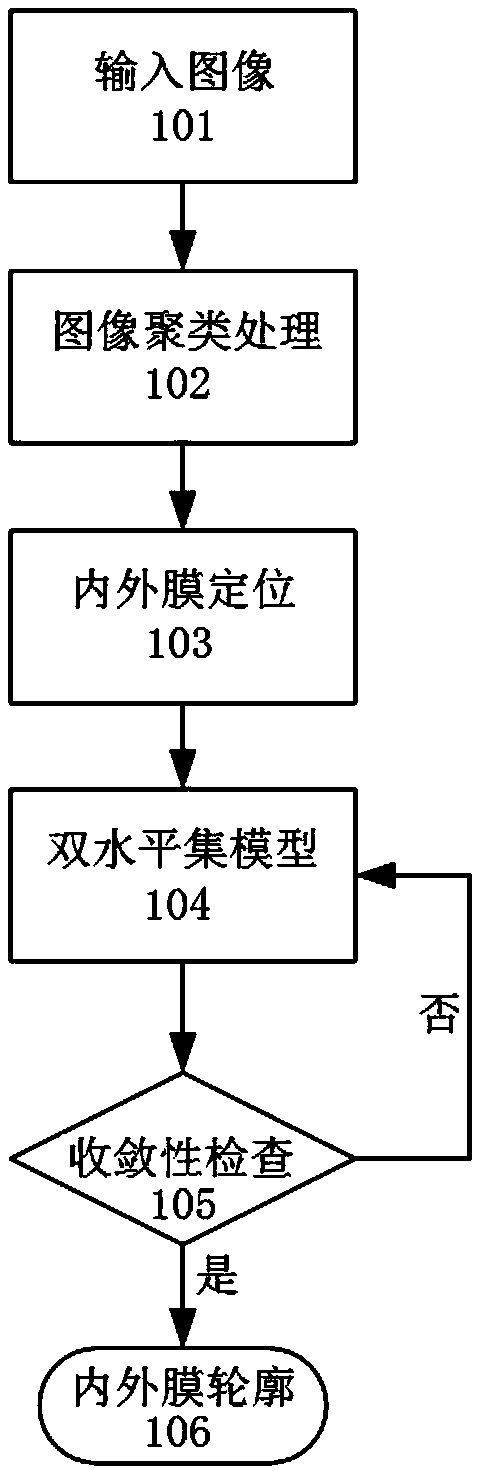
[0103] The present invention will be further described below in conjunction with accompanying drawing. see Figures 1 to 10g , a method for automatically segmenting the inner and outer membranes of the left ventricle, the specific steps are as follows:

[0104] 1) Select the left ventricular MRI image to be processed (such as figure 2 ), and input image 101;

[0105] 2) Image clustering processing 102, select the optimized Mean Shift clustering algorithm to preprocess the left ventricle MRI image:

[0106] Resolve blurry borders:

[0107] Due to the close distance between the border of the left ventricle and the right ventricle of the image, the contrast of the underlying visual features of the image is small. In order to better obtain the initial detection position, the image is first processed with fuzzy sets. Since the Mean Shift algorithm is mainly used in cluster analysis in computer vision and image processing, it is a non-parametric feature space analysis technique...


PUM


Abstract

The invention discloses a method for automatically segmenting left ventricular inner and outer membranes, which comprises the following steps: 1) selecting an image to be processed; 2) preprocessing the left ventricular image by using the optimized Mean shift clustering algorithm; 3) using the improved hough transform circle detection algorithm to locate the left heart lumen and obtain the initialcontour of inner and outer membrane segmentation; 4) segmenting inner and outer membranes of the image by a two-level model; 5) checking the convergence of the energy function formula of the two-level set model; 6) obtaining a segmentation effect map of the segmented inner and outer membrane contour. The invention solves the problem of artificially setting the initial position of the segmentation, improves the segmentation precision of the inn and outer membrane, reduces the time of the model segmentation of the inner and outer membrane, and obtains the inner and outer membrane contour without edge leakage and with good regularity, which conforms to the clinical definition.

Description

technical field [0001] The invention relates to medical image processing technology, in particular to a method for automatically dividing the inner and outer membranes of the left ventricle. Background technique [0002] In recent years, the increasing number of deaths from heart disease has triggered people's deeper exploration of the heart. Accurate extraction of the left ventricular epicardium is an important basis for further research on the left ventricle of the heart. In terms of clinical application, the accurate segmentation of the left ventricular epicardium can provide some important parameters of left ventricular function, which is helpful for doctors to quantitatively analyze whether the left ventricular function is abnormal; in terms of scientific research, the segmentation of the left ventricle is the basis for three-dimensional modeling of the left ventricle. The amount of image data in a heart vibration cycle is large. If you want to obtain objective and acc...

Claims


Application Information

Patent Timeline
IPC IPC(8): G06T7/11G06T7/12G06T7/13G06T7/136G06K9/62
CPCG06T7/11G06T7/12G06T7/13G06T7/136G06T2207/10088G06T2207/20061G06T2207/30048G06F18/2321
Inventor 李军华李林
Owner NANCHANG HANGKONG UNIVERSITY
Why Eureka
Try Eureka