An automatic identification method of blood flow velocity waveform based on ultrasound image

A technology of blood flow velocity and ultrasound image, which is applied in image analysis, image enhancement, image data processing, etc., to save time and simplify the extraction process

Active Publication Date: 2022-03-22
BEIJING UNIV OF TECH
View PDF5 Cites 0 Cited by
  • Summary
  • Abstract
  • Description
  • Claims
  • Application Information

AI Technical Summary

Problems solved by technology

The waveform can be output after eliminating interference such as digital borders

Method used


Image

  • An automatic identification method of blood flow velocity waveform based on ultrasound image
  • An automatic identification method of blood flow velocity waveform based on ultrasound image
  • An automatic identification method of blood flow velocity waveform based on ultrasound image

Examples


Embodiment 1

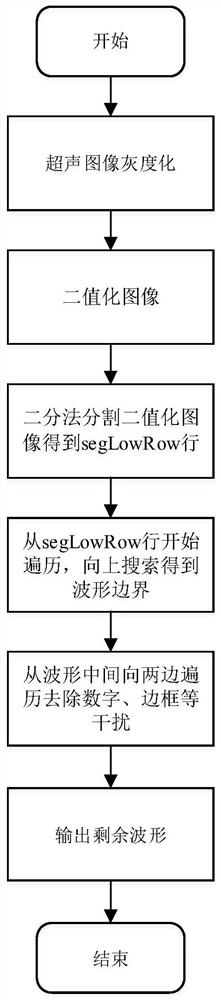
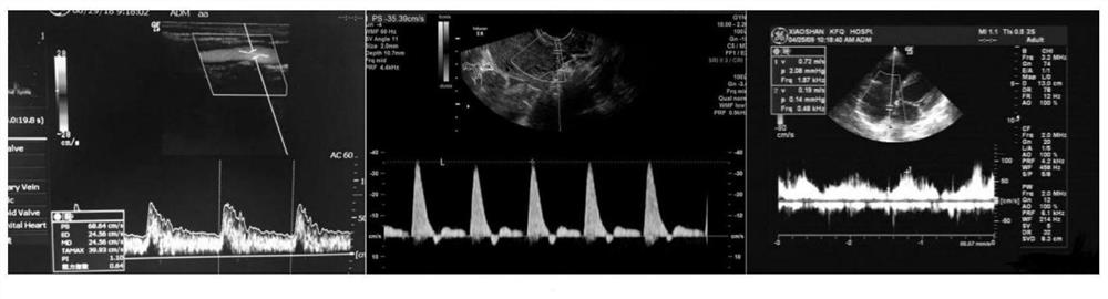
[0029] Step A1: Select three different ultrasonic images containing blood flow velocity waveforms, and grayscale the ultrasonic images to obtain a grayscale image;

[0030] Step A2: carry out binarization processing to grayscale image and obtain binarized image;

[0031] Due to the high gray value of the blood flow velocity waveform in most ultrasound images, the binarization threshold was set to 100 based on the experimental experience of three kinds of ultrasound images.

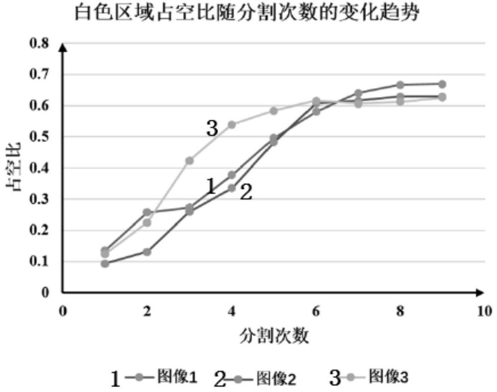
[0032] Step A3: using the dichotomy method to perform multiple horizontal segmentations on the binarized image to obtain the area of ​​high gray value below the blood flow velocity waveform;

[0033] For most ultrasound images, when the white area with high gray value under the blood flow velocity waveform is segmented, the duty cycle growth rate of the white area will decrease. The successive dichotomous segmentation of three different ultrasound images, the duty ratio of the respective white areas ima...


PUM


Abstract

The invention relates to an automatic identification method of blood flow velocity waveform based on an ultrasonic image, which belongs to the field of image processing algorithms. The method is based on the collected or photographed clinical ultrasound images containing blood flow velocity waveforms, and performs grayscale and binarization preprocessing on the images; The area of ​​high gray value below the waveform; traverse upwards from the last line of the segmented area as a whole, and when the maximum gray gradient is experienced and there is no high-brightness gray in a certain length of area that continues upward, the boundary of the waveform can be identified. That is, the blood flow velocity waveform; finally, it traverses from the middle position of the waveform to both ends to remove the non-waveform numbers, borders and other interferences, and output the digital waveform. The invention realizes the automatic identification and digital output of the blood flow velocity waveform in the image, and lays a foundation for the analysis of the blood flow velocity waveform when the waveform source data of the ultrasonic equipment cannot be obtained.

Description

technical field [0001] The invention belongs to the field of image processing algorithms, and relates to an automatic identification method of blood flow velocity waveform based on ultrasonic images. Background technique [0002] Ultrasound is a routine blood flow velocity detection method in clinic, and it has a wide range of application value in the diagnosis of human diseases. The blood flow velocity waveform detected by ultrasound equipment contains a wealth of information related to the disease. Whether in clinical real-time observation or in the follow-up analysis of the disease, the blood flow waveform has important reference significance. The amplitude and shape of the blood flow waveform can be observed and analyzed manually in real time, but apart from these obvious features, the waveform may contain disease-related information that cannot be directly observed. In the subsequent analysis of diseases, this information can be discovered through digital signal proces...

Claims


Application Information

Patent Timeline
Patent Type & Authority Patents(China)
IPC IPC(8): G06T7/00G06T7/11
CPCG06T7/0012G06T7/11G06T2207/10132G06T2207/30104
Inventor 刘有军李鲍王文馨毛伯䶮
Owner BEIJING UNIV OF TECH
Why Eureka
Try Eureka