
Ventricular region segmentation model training method and device and template image determining method and device

A region segmentation and template image technology, applied in the field of image processing, can solve the problems of affecting the mapping effect of the MCA blood supply area, low registration accuracy, and difficulty in accurately determining the blood supply of the MCA, etc.

Pending Publication Date: 2022-02-18
沈阳东软智能医疗科技研究院有限公司
View PDF0 Cites 0 Cited by
  • Summary
  • Abstract
  • Description
  • Claims
  • Application Information

AI Technical Summary

Problems solved by technology

When a variety of different brain CT images are registered based on a fixed template image and the MCA brain atlas image corresponding to the template image, the registration difficulty between the brain CT image and the template image will increase, resulting in low registration accuracy. Affects the mapping of the MCA blood supply, making it difficult to accurately determine the blood supply of the MCA

Method used


Image

  • Ventricular region segmentation model training method and device and template image determining method and device
  • Ventricular region segmentation model training method and device and template image determining method and device
  • Ventricular region segmentation model training method and device and template image determining method and device

Examples


specific Embodiment approach

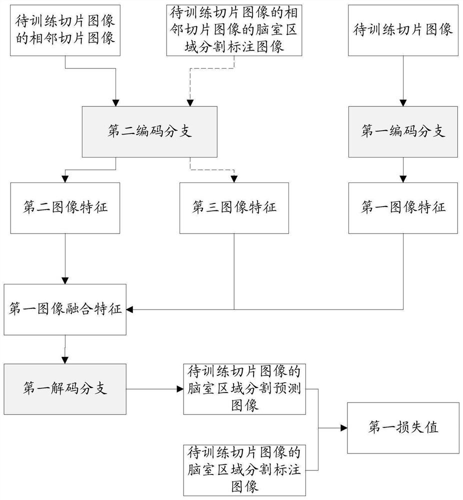
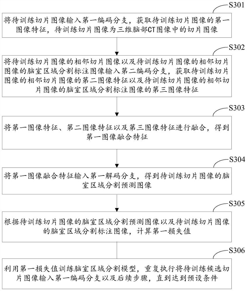
[0142] During specific implementation, the candidate slice images are firstly determined from the three-dimensional brain CT images. The candidate slice images are all slice images or partial slice images in the three-dimensional brain CT image. If it is all slice images, the efficiency of determining typical slice images from all slice images is relatively low. To this end, the embodiment of the present application provides a specific implementation method for determining candidate slice images from three-dimensional brain CT images, including:

[0143] A1: The three-dimensional brain CT image is binarized according to the CT threshold to obtain the brain region, and the slice image with the largest brain region area in each slice image of the three-dimensional brain CT image is determined as the central slice image.

[0144] The three-dimensional brain CT image is binarized, and the background area and brain area in the three-dimensional brain CT image are divided by CT thr...


PUM


Abstract

The embodiment of the invention discloses a training method of a ventricular region segmentation model, and a method, device and equipment for determining a template image of a brain CT image. The method comprises steps of determining candidate slice images from a three-dimensional brain CT image of a patient, determining a ventricle region and a brain parenchyma region of each candidate slice image, and calculating the area ratio of the ventricle region to the brain parenchyma region of each candidate slice image, and determining the candidate slice image corresponding to the maximum value in the area ratio as a typical slice image, and selecting a template image matched with the area ratio of the ventricle region to the brain parenchyma region of the typical slice image from a template image group corresponding to the age of the patient, and taking the template image as a template image corresponding to the three-dimensional brain CT image. Based on the area ratio of the ventricle region to the brain parenchyma region of the typical slice image, the matching degree of the template image selected from the template image group corresponding to the age of the patient and the three-dimensional brain CT image is high, and the registration difficulty of the subsequent brain CT image and the template image can be reduced.

Description

technical field [0001] The present application relates to the field of image processing, in particular to a training method for a segmentation model of a ventricular region, a method, a device and equipment for determining a template image of a brain CT image. Background technique [0002] Ischemic stroke is a major disease that threatens human health. ASPECTS (Alberta Stoke Program Early CT Score, Alberta Stroke Program Early CT Score) can be used to judge the blood supply of multiple blood supply regions in the MCA (Middle Cerebral Artery, Middle Cerebral Artery) of patients with ischemic stroke, which can help doctors understand the blood supply of patients . [0003] MCA blood supply area mapping is the core link in ASPECTS. MCA blood supply area mapping refers to registering the brain CT image and the template image corresponding to the brain CT image to obtain the deformation field between the brain CT image and the template image corresponding to the brain CT image....

Claims


Application Information

Patent Timeline
IPC IPC(8): G06T7/00G06T7/11G06T7/136G06T7/62G06N3/04G06N3/08G06K9/62G06V10/44G06V10/764G06V10/82
CPCG06T7/0012G06T7/11G06T7/62G06T7/136G06N3/04G06N3/08G06T2207/10081G06T2207/30016G06T2207/20081G06F18/2321
Inventor 杨明雷袁红美钱山贾晓甜
Owner 沈阳东软智能医疗科技研究院有限公司
Learn More