Tomosynthetic image reconstruction method, and diagnostic device operating according to the method

a tomosynthetic and image reconstruction technology, applied in the field of tomosynthetic image reconstruction methods, can solve the problems of difficult detection, inability to achieve the image quality known from computed tomography (ct), and reconstruction algorithms, as are known from tomography, can not be used for tomosynthetic reconstruction without any problems, and achieve the effect of minimal processing effor

Active Publication Date: 2007-09-20
SIEMENS HEALTHCARE GMBH
View PDF11 Cites 6 Cited by
  • Summary
  • Abstract
  • Description
  • Claims
  • Application Information

AI Technical Summary

Benefits of technology

[0011]An object of the invention is to provide a tomosynthesis image reconstruction method, especially for mammography, with which, using minimal processing effort, a 3D x-ray image can be ge

Problems solved by technology

Since in such a projection the tissue layers lying behind each other in the direction of the x-ray beam are overlaid, heavily-absorbent beneficial structures can overlay a harmful tumor and render such detectability more difficult.
Because of the incomplete scanning, i.e. the projections are only available from a restricted angular range, a reconstruction of a 3D x-ray image is only p

Method used


Image

  • Tomosynthetic image reconstruction method, and diagnostic device operating according to the method
  • Tomosynthetic image reconstruction method, and diagnostic device operating according to the method
  • Tomosynthetic image reconstruction method, and diagnostic device operating according to the method

Examples


Embodiment Construction

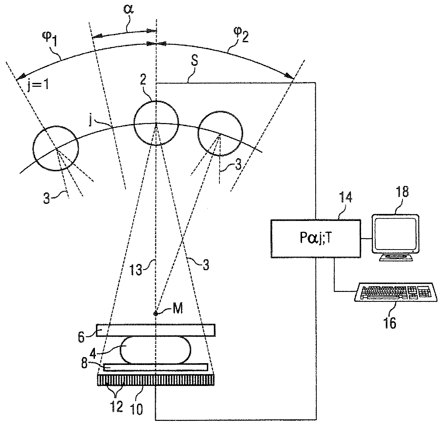
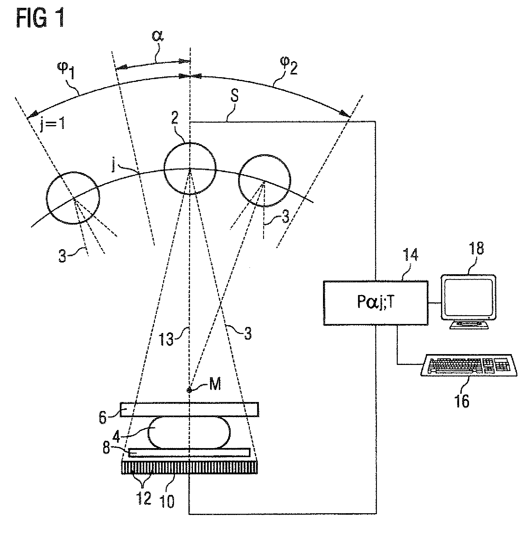
[0033]According to FIG. 1 the device, in the exemplary embodiment a mammography device, has an x-ray tube 2 for generating x-rays 3 that pass through an object under examination 4. The object under examination 4 is a female breast which is held between a compression plate 6 and a support plate 8. The x-rays passing through the object under examination 4, the compression plate 6 and the support plate 8 are received by a wide-area digital x-ray detector 10 which is formed by a number of individual detectors 12 arranged in a matrix-shaped array, and of which the receive surface 11 is arranged in parallel to the plates 6, 8.

[0034]The x-ray tube 2 is arranged to enable its location to be changed in a restricted area in relation to the object under examination, and can for example within a restricted angular range φ1,φ2 be pivoted around an axis M perpendicular to the recording plane into different angular positions j=1 . . . n, so that individual images of the object under examination 4 ...


PUM


Abstract

In a tomosynthetic image reconstruction method and diagnostic device operating with such a method, a tomosynthetic 3D x-ray image is reconstructed by a discrete filtered back projection from a number of individual digital projection data recorded from different project angles within a restricted angular range, in which at least one filtering is performed with a convolution kernel that, in the local area outside of its central value, corresponds to an exponential function.

Description

BACKGROUND OF THE INVENTION[0001]1. Field of the Invention[0002]The invention relates to a tomosynthetic image reconstruction method, especially suitable for mammography, in which a tomosynthetic 3D x-ray image is assembled from individual digital images recorded from a number of different projection angles. In addition the invention relates to a diagnostic device operating with such a method.[0003]2. Description of the Prior Art[0004]Mammography involves x-ray examination of the female breast for the purpose of detecting tumors at the earliest possible stage. By constantly improving mammography methods an attempt is made to generate x-ray images supplying a high level of information, in order to distinguish beneficial from harmful changes and to reduce the number of incorrect findings, i.e. the number of suspicious findings that are caused by non-harmful changes, and the number of the undiscovered harmful tumors. In conventional x-ray mammography in this case a two-dimensional sing...

Claims


Application Information

Patent Timeline
IPC IPC(8): H05G1/60A61B6/00G01N23/00G21K1/12
CPCG06T11/006G06T2211/421
Inventor BARTH, KARLHAERER, WOLFGANGMERTELMEIER, THOMASWIESENT, KARL
Owner SIEMENS HEALTHCARE GMBH
Why Eureka
Try Eureka