Rapid exchange fna biopsy device with diagnostic and therapeutic capabilities

a biopsy device and fna technology, applied in the field of needle biopsy devices, can solve the problems of cumbersome means of attaching a device to an echoendoscope, inability to examine nearby organs, and inability to perform ovulation diagnostics, etc., to facilitate the efficient delivery of desired agents, facilitate the efficient delivery of fiducial markers, and facilitate the efficient delivery of implantable biomarkers

Inactive Publication Date: 2011-08-04
TYCO HEALTHCARE GRP LP
View PDF114 Cites 66 Cited by
  • Summary
  • Abstract
  • Description
  • Claims
  • Application Information

AI Technical Summary

Benefits of technology

[0017]As will be described later, the devices of the invention can be configured to deliver agents in the form of liquids, gels, glues, seeds, pellets, encapsulated liquids or encapsulated gels, to desired locations in the gastrointestinal and respiratory systems to treat various cancerous or other tumors. For example, the devices of the invention can be used to facilitate the efficient delivery of a desired agent, such as chemotherapeutic agents, sclerosing agents, tumor necrosing factors, growth factors (pharma and bio agents), and / or imaging agents to desired sites in the gastrointestinal or respiratory tracts, to provide a localized therapeutic effect in the treatment of benign and malignant tumors. The devices of the invention can also be used to deliver radiation therapy directly to tumors diagnosed in the respiratory and gastrointestinal tracts to aid in the treatment of cancer. The desired agent can be delivered to the target site by introducing the desired agent in a liquid, gel or glue form through one or more of the ports in the needle housing member. Alternatively, the desired agent can be delivered to the target site using a needle pre-loaded with the desired agent in an encapsulated form, or pellet or seed form.
[0018]The devices of the invention can also be used to facilitate the efficient delivery of fiducial markers to desired sites in the gastrointestinal or respiratory tracts, to provide “landmarks” in the anatomy to aid in directed radiotherapy or other therapy.
[0019]The devices of the invention can also be used to facilitate the efficient delivery of implantable biomarkers that may be used as part of an image guided system in conjunction with Fine Needle Aspiration (FNA) using ultrasound guided imaging techniques to desired sites in the gastrointestinal or respiratory tracts, to provide an identifying position for the delivery of subsequent therapy during patient treatment.
[0025]In each of the aforementioned embodiments, the FNA devices of the invention can further include a strain relief to stabilize the needle by providing a smooth transition and bend radius for the needle housing member upon insertion and removal from the proximal handle member.

Problems solved by technology

Although a high frequency ultrasound can produce a higher image quality, high frequency ultrasounds do not penetrate organ walls as well as lower frequency ultrasound.
As a result, the examination of the nearby organs is not possible.
These device, however, suffer from several drawbacks.
For example, the means of attaching a device to an echoendoscope is cumbersome.
In addition, these devices provide sub-optimal ergonomics of use.
The cumulative actions required by a clinician result in significantly drawn out procedures.
Further, needles commonly kink or deform during removal from a device causing numerous delays and failures.
Moreover, multiple passes per procedure are required, which prolong the procedure and result in a clinician needing to reconfirm the location of a needle relative to a desired aspiration site with each new pass.
However, in the current field of therapeutic endoscopy and pulmonology, the ability of the physician to effectively and efficiently treat known metastases, strictures, tumors etc., in the gastrointestinal tract, the endobronchial tract, and peripheral areas, is greatly hampered by the lack of available technology to deliver diagnostic aids, reagents and other therapeutic means to the desired treatment site.

Method used


Image

  • Rapid exchange fna biopsy device with diagnostic and therapeutic capabilities
  • Rapid exchange fna biopsy device with diagnostic and therapeutic capabilities
  • Rapid exchange fna biopsy device with diagnostic and therapeutic capabilities

Examples


Embodiment Construction

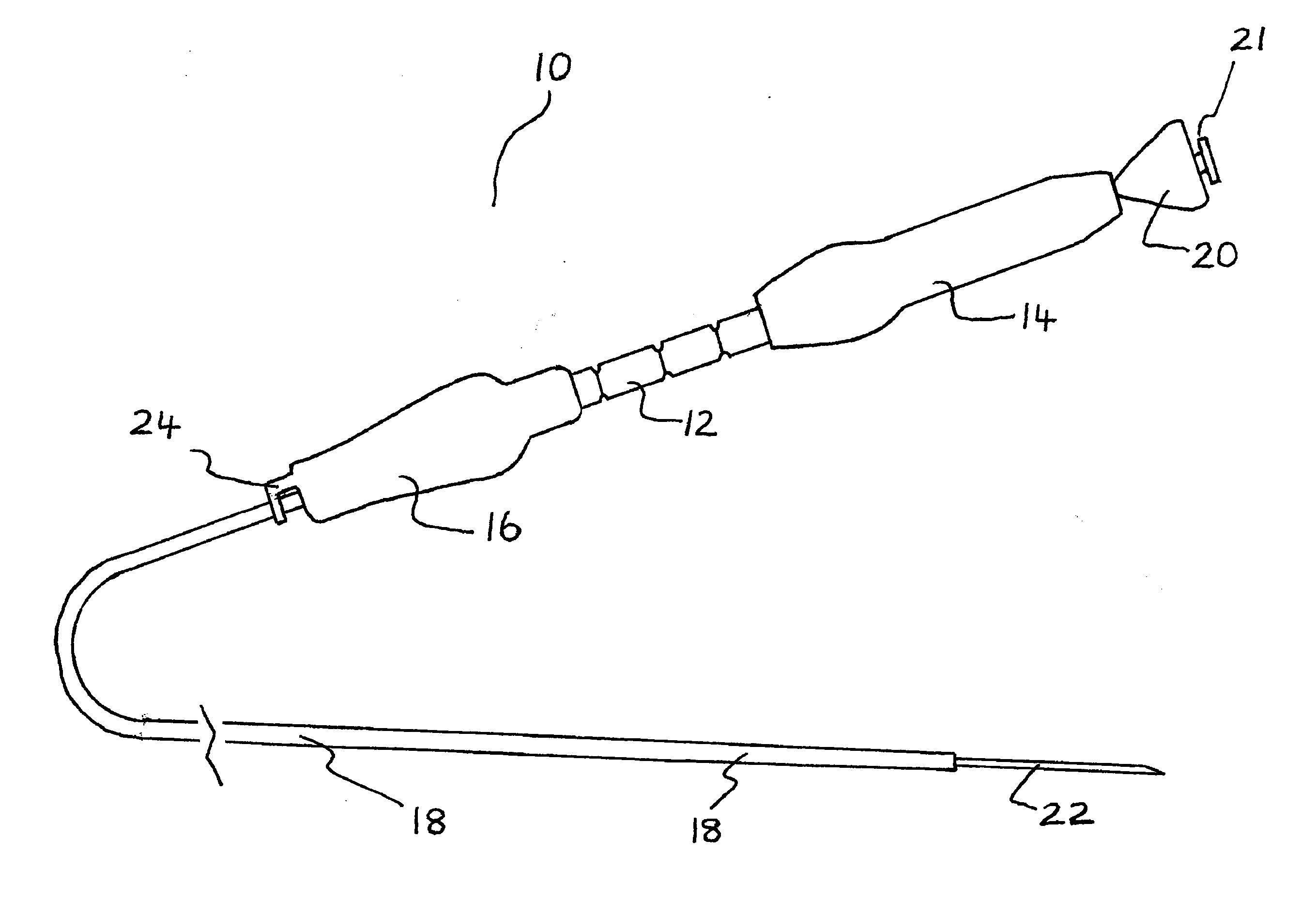
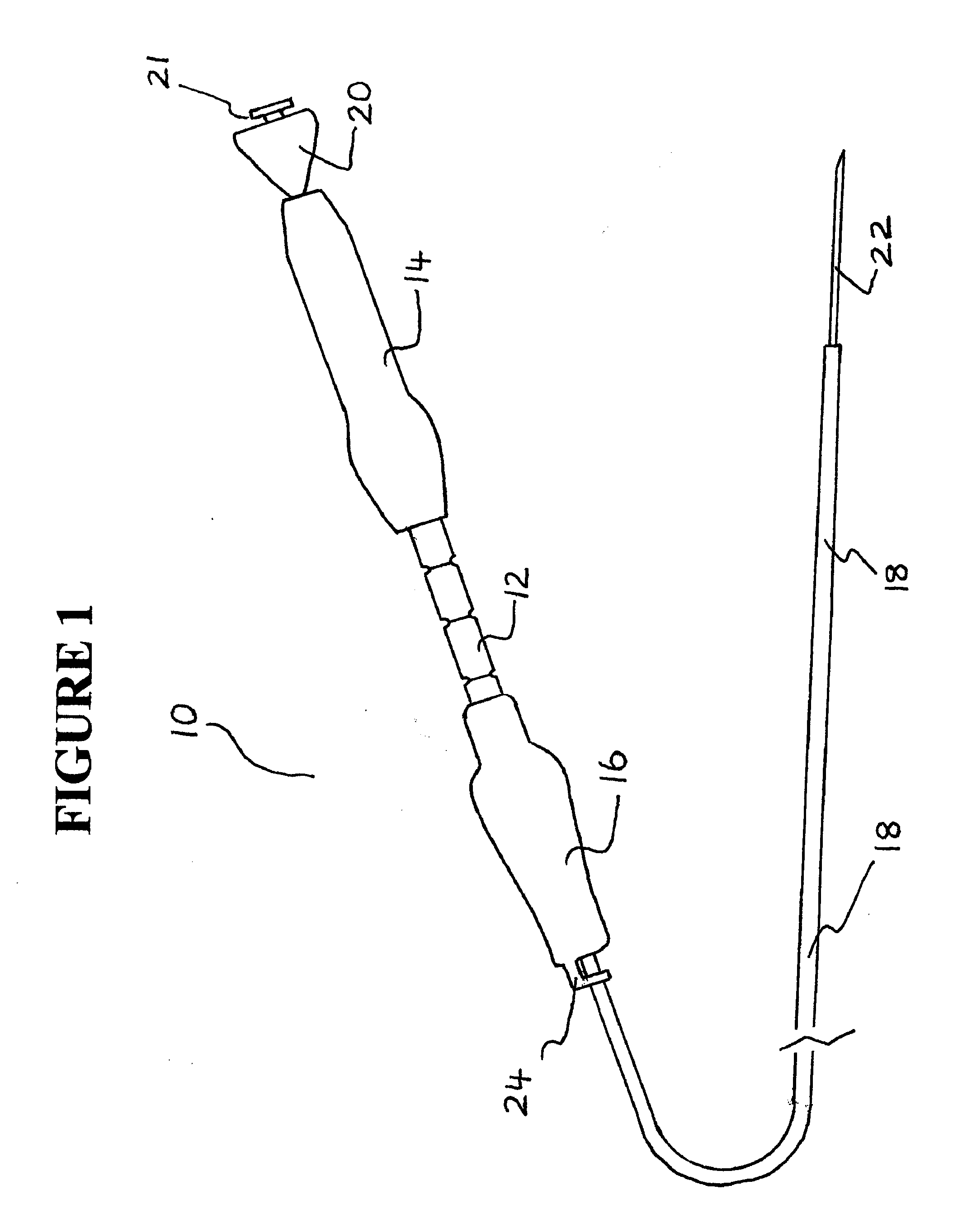
[0065]The exemplary embodiments of the needle biopsy device and methods of operation disclosed are discussed in terms of needle biopsy devices for collecting tissue, fluid, and cell samples from a body in conjunction with an endoscopic ultrasound or endoscopic bronchial ultrasound. It is envisioned that the present disclosure, however, finds application to a wide variety of biopsy devices for the collection of samples from a subject. It is also envisioned that the present disclosure may be employed for collection of body fluids including those employed during procedures relating to phlebotomy, digestive, intestinal, urinary, veterinary, etc. It is contemplated that the needle biopsy device may be utilized with other needle biopsy applications including, but not limited to, fluid collection, catheters, catheter introducers, spinal and epidural biopsy, aphaeresis, dialysis, etc. Suitable needle biopsy devices are described in U.S. patent application Ser. No. 12 / 243,367 (published as U...


PUM


Abstract

A device for needle biopsy and delivery of a diagnostic or therapeutic agent is presented. The device includes a handle member having proximal and distal portions. A proximal handle member is disposed to the proximal portion of the handle member and a distal handle member is disposed to the distal portion of the handle member. A sheath lumen is disposed within the handle member and extends from the distal portion of the handle member. A needle housing member is partially disposed to the proximal portion of the handle member and a needle is disposed within the sheath lumen. The needle housing member includes one or more ports for introducing an agent or a device into the housing member. The needle can include an agent or a device disposed at a distal portion thereof.

Description

CROSS REFERENCE TO RELATED APPLICATIONS[0001]This application is a continuation-in-part application of U.S. patent application Ser. No. 12 / 243,367, filed on Oct. 1, 2008, and a continuation-in-part of U.S. patent application Ser. No. 12 / 607,636, filed on Oct. 28, 2009, which in turn claims the benefit of U.S. Provisional Application Ser. No. 61 / 117,966, filed on Nov. 26, 2008, and U.S. Provisional Application Ser. No. 61 / 152,741 filed on Feb. 16, 2009. The contents of each of these applications are incorporated by reference herein in their entireties. This application further claims priority under 35 U.S.C. 119(e) to U.S. Provisional Application No. 61 / 305,304, filed on Feb. 17, 2010, and U.S. Provisional Application No. 61 / 305,396, filed on Feb. 17, 2010, the contents of which are incorporated by reference herein in their entireties.FIELD OF THE INVENTION[0002]The present invention generally relates to biopsy devices, and more particularly, to needle biopsy devices for collecting t...

Claims


Application Information

Patent Timeline
Patent Type & Authority Applications(United States)
IPC IPC(8): A61B10/02
CPCA61B10/04A61B17/1114A61B2010/045A61B2017/1139A61F2002/041A61B2019/5487A61F2/848A61F2/88A61F2/966A61B2017/347A61B2090/3987
Inventor MCWEENEY, JOHN
Owner TYCO HEALTHCARE GRP LP
Why Eureka
Try Eureka