Dual energy imaging using optically coupled digital radiography system

a digital radiography and optically coupled technology, applied in the field of digital radiography, can solve the problems of less than perfect separation of bone and soft tissue components, laborious and time-consuming digital imaging using phosphor-based receptor plates,

Inactive Publication Date: 2006-03-07
1370509 ALBERTA
View PDF27 Cites 33 Cited by
  • Summary
  • Abstract
  • Description
  • Claims
  • Application Information

AI Technical Summary

Problems solved by technology

Digital imaging using phosphor-based receptor plates is laborious and time intensive as technologists typically must carry the plates to a reader and wait for the reader to energize the plates and record light flashes that correspond to the energy imparted by the x-rays that struck the plates.
Because this technique involves multiple sequential exposures, the time delay between exposures tends to cause misregistration resulting in a less-than-perfect separation of the bone and soft tissue components.

Method used


Image

  • Dual energy imaging using optically coupled digital radiography system
  • Dual energy imaging using optically coupled digital radiography system
  • Dual energy imaging using optically coupled digital radiography system

Examples


Embodiment Construction

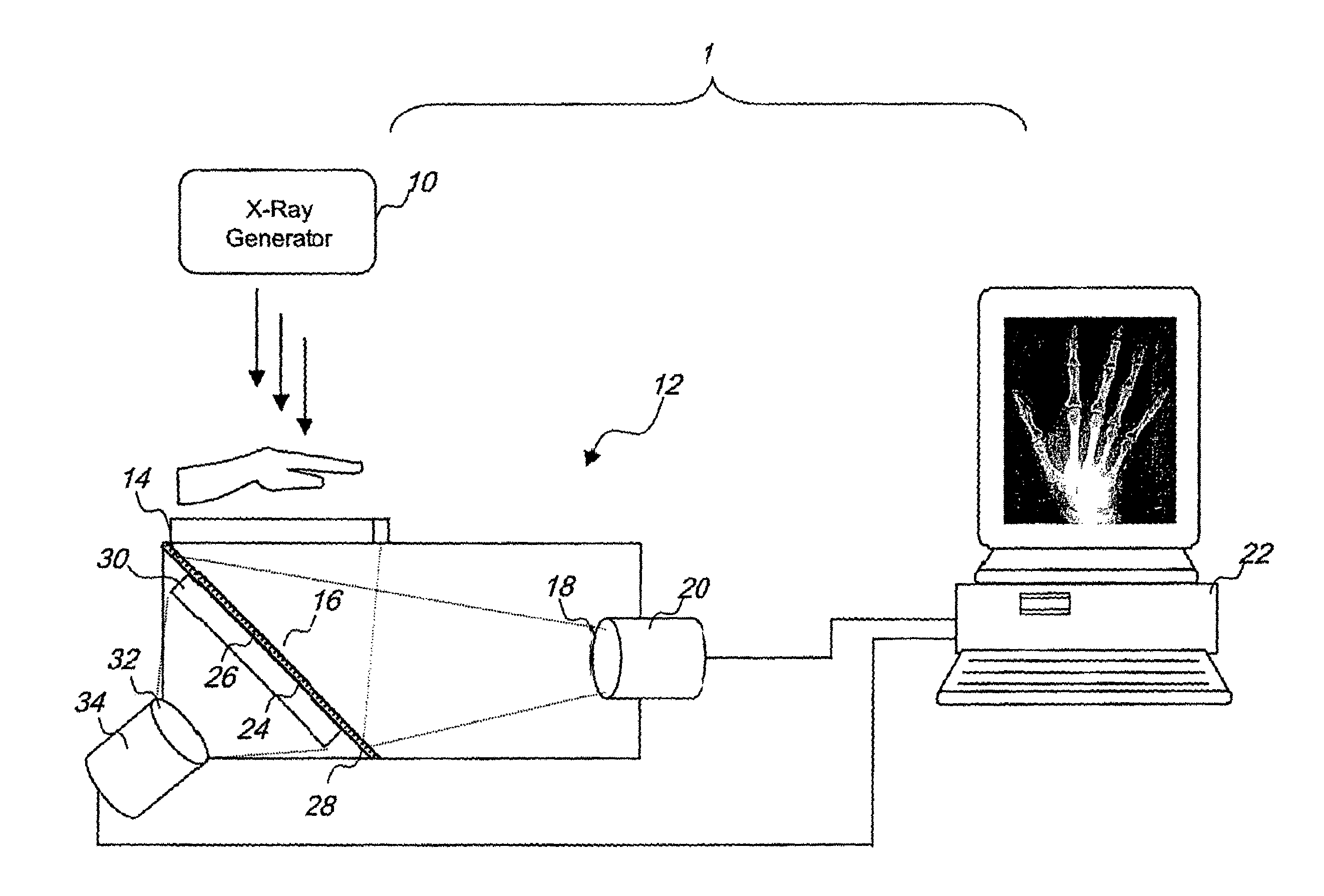
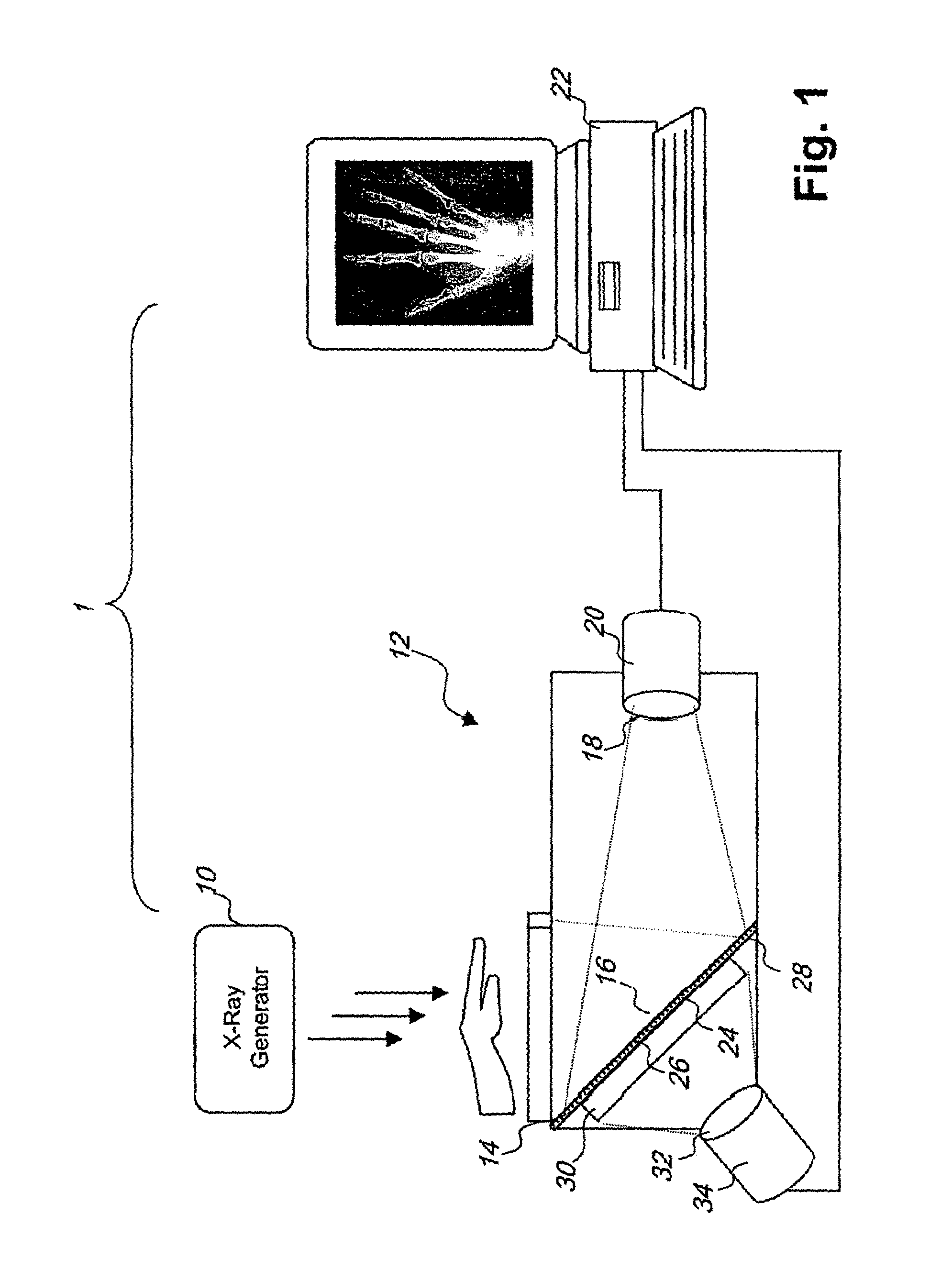
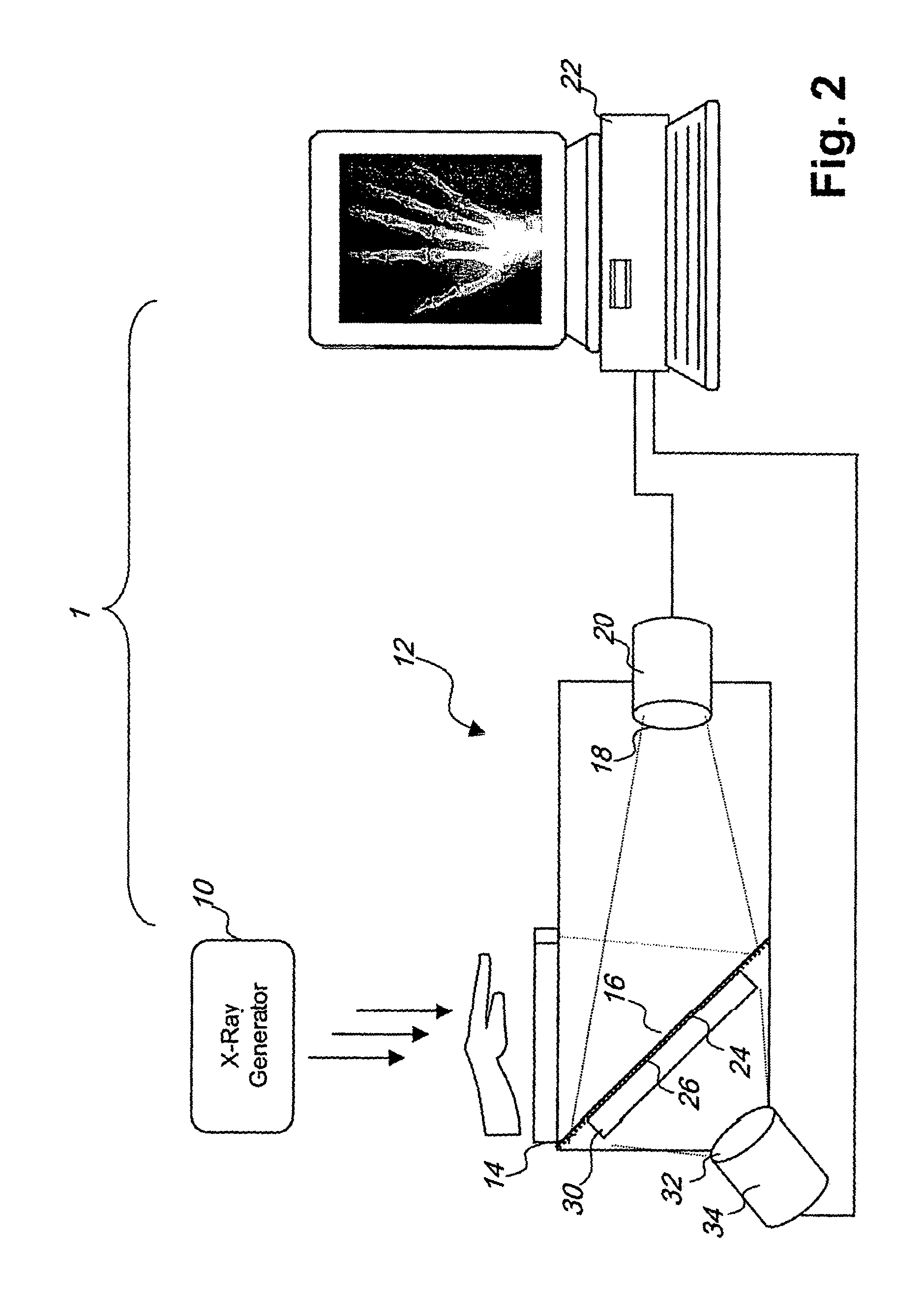
[0028]Referring to FIG. 1 and according to one embodiment of the invention, an optically-coupled CCD-based DR system 1 is provided for taking digital x-ray images of a subject, such as a human patient, for clinical diagnostic purposes.

[0029]The system 1 is operable to simultaneously obtain two distinct images of a subject, each of which represents a different x-ray energy spectrum. The two images can be algebraically combined in various ways during image processing, such that anatomical features can be separated from one another to provide a clearer view of certain features of underlying structures. In particular, one image can be algebraically combined with another to produce a third image that enhances the bone structure or muscle tissue in the subject.

[0030]In particular, the two different-energy images obtained by the system 1 can be processed to produce a third image showing only bone or only soft tissue. The process uses a set of intensity reference tables provided for each sc...


PUM


Abstract

This invention relates to an optically coupled digital radiography method and apparatus for simultaneously obtaining two distinct images of the same subject, each of which represents a different x-ray energy spectrum. The two images may be combined in various ways such that anatomical features may be separated from one another to provide a clearer view of those features or of underlying structures. The two different images are obtained using a pair of scintillators separated by an x-ray filter that attenuates part of the x-ray spectrum of an x-ray exposure such that the first and second scintillators receive a different energy spectrum of the same x-ray exposure. Alternatively, the two different images can be obtained without a filter and with two scintillators made of different fluorescing materials that react differently to the same x-ray exposure.

Description

FIELD OF THE INVENTION[0001]The present invention is directed generally to digital radiography, and in particular to an optically-coupled digital radiography system that can simultaneously acquire two images with different x-ray energy spectra for the purpose of producing separable bone and soft tissue images.BACKGROUND OF THE INVENTION[0002]For over a hundred years photographic films have been used to capture and display x-rays for diagnostic purposes. In recent years, digital radiography (DR) has become increasingly popular. DR refers to the application of digital equipment and image processing techniques to projection radiography. Digitally recorded x-rays are superior to those recorded with photographic film due to tile greater dynamic range offered by a digital recording system. Furthermore, computer image processing techniques provide a wealth of capabilities to study otherwise obscured details within the image.[0003]One type of DR imaging device is an optically-coupled charge...

Claims


Application Information

Patent Timeline
Patent Type & Authority Patents(United States)
IPC IPC(8): H05G1/64G01T1/20A61B6/00G01T1/208G01T1/36
CPCG01T1/362A61B6/505A61B6/482A61B6/4035
Inventor WINSOR, ROBIN
Owner 1370509 ALBERTA
Why Eureka
Try Eureka