Encapsulated medical device and method of examining, curing, and treating internal region of body cavity using encapsulated medical device

a medical device and encapsulation technology, applied in the field of encapsulated medical devices, can solve the problems of difficult to keep the endoscope inserted therein for a prolonged period of time, balloons coming into close contact with the internal surface of the body cavity to block the lumen, and concave parts hardly allow fluid such as gas or humor to flow forward or backward in the lumen, so as to achieve the effect of not deteriorating the ease of swallowing of the encapsul

Inactive Publication Date: 2006-08-01
OLYMPUS CORP
View PDF4 Cites 151 Cited by
  • Summary
  • Abstract
  • Description
  • Claims
  • Application Information

AI Technical Summary

Benefits of technology

[0013]Accordingly, an object of the present invention is to provide an encapsulated medical device and a method of examining, curing, or treating an internal region of a body cavity using the encapsulated medical device. Herein, the encapsulated medical device allows a fluid such as

Problems solved by technology

It is however hard to keep the endoscope inserted therein for a prolonged period of time.
However, in the peroral endoscope described in the Japanese Unexamined Patent Application Publication No. 2000-342522 has a drawback that the balloon comes into close contact with the internal surfac

Method used


Image

  • Encapsulated medical device and method of examining, curing, and treating internal region of body cavity using encapsulated medical device
  • Encapsulated medical device and method of examining, curing, and treating internal region of body cavity using encapsulated medical device
  • Encapsulated medical device and method of examining, curing, and treating internal region of body cavity using encapsulated medical device

Examples


first embodiment

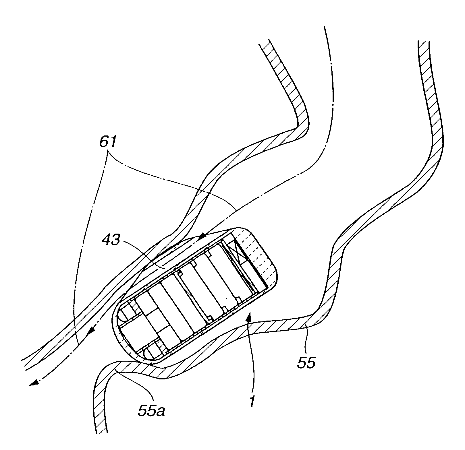
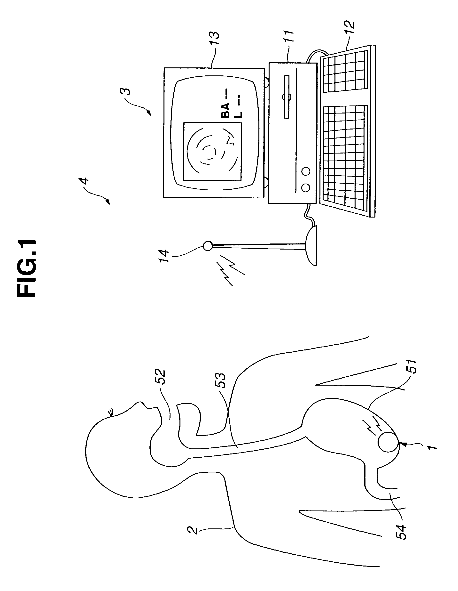
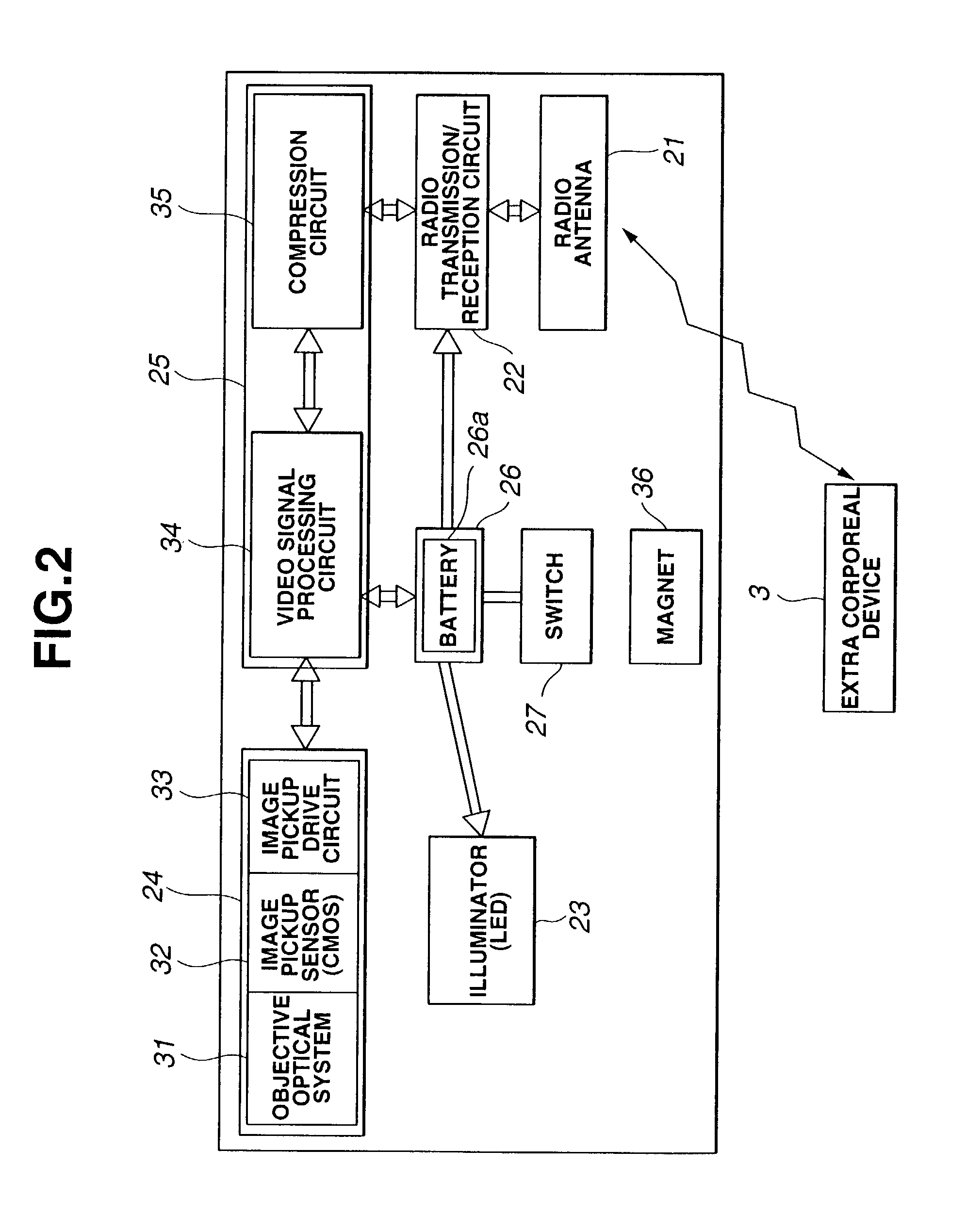
[0062]FIG. 1 to FIG. 12B are concerned with a first embodiment of the present invention. FIG. 1 shows the overall configuration of a medical system including the first embodiment of the present invention. FIG. 2 is a circuit block diagram showing the encapsulated medical device in accordance with the first embodiment of the present invention. FIG. 3A is a sectional view showing the components of the encapsulated medical device shown in FIG. 2. FIG. 3B shows the appearance of the front part of the encapsulated medical device shown in FIG. 3A. FIG. 3C shows the appearance of the-back part of the encapsulated medical device shown in FIG. 3A. FIG. 4 is a sectional view showing a scene where the encapsulated medical device shown in FIG. 3A to FIG. 3C is passing through the esophagus. FIG. 5 is a sectional view showing a scene where the encapsulated medical device shown in FIG. 3A to FIG. 3C has come to a standstill in a stenosed part of the small intestine. FIG. 6 is a sectional view of ...

second embodiment

[0121]FIG. 13A to FIG. 15 are concerned with a second embodiment of the present invention. FIG. 13A is a sectional view showing the components of an encapsulated medical device in accordance with the second embodiment of the present invention. FIG. 13B is an A—A sectional view of the encapsulated medical device shown in FIG. 13A. FIG. 13C is a B—B sectional view of the encapsulated medical device shown in FIG. 13A. FIG. 13D is a C—C sectional view of the encapsulated medical device shown in FIG. 13A. FIG. 14 is a D—D sectional view of the encapsulated medical device shown in Fig. B. FIG. 15 is a sectional view showing a scene where the encapsulated medical device shown in FIG. 13A to FIG. 14 has come to a standstill in a stenosed part of the small intestine.

[0122]In the aforesaid first embodiment, the encapsulated medical device is formed with one capsule body. In the second embodiment, a capsule body is divided into a distal hard member and a back hard member. The distal hard membe...

third embodiment

[0133]FIG. 16A to FIG. 21B are concerned with a third embodiment of the present invention. FIG. 16A is a sectional view showing the components of an encapsulated medical device in accordance with the third embodiment of the present invention. FIG. 16B is an E—E sectional view of the encapsulated medical device shown in FIG. 16A. FIG. 17 is an explanatory diagram showing an encapsulated medical device of a variant that has a capsule body thereof covered with a transparent member that is an armor member. FIG. 18 is an explanatory diagram showing a scene where the encapsulated medical device shown in FIG. 17 has come to a standstill in a stenosed part of the small intestine. FIG. 19 is an explanatory diagram showing an encapsulated medical device of a variant that has a net-like mesh cover (mesh jacket) freely detachably attached to a capsule body thereof. FIG. 20 is an explanatory diagram showing a scene where the encapsulated medical device shown in FIG. 19 has come to a standstill i...


PUM


Abstract

An encapsulated medical device includes a radio antenna through which the encapsulated medical device transmits or receives a radio wave to or from an extracorporeal device. The encapsulated medical device is passed through the lumen of a body cavity in order to examine, cure, or treat an internal region of the body cavity under the control of the extracorporeal device. A capsule body included in the encapsulated medical device has an extended portion formed at an upper end of one axis of a cross section of the capsule body cut at right angle to the longitudinal axis thereof. The capsule body has a linkage hole bored in the extended portion thereof to allow a fluid such as a gas or humor to flow into forward and backward parts of the lumen through the hole.

Description

[0001]This application claims the benefit of Japanese Application No. 2001-297703 filed in Japan on Sep. 27, 2001, the contents of which are incorporated by reference.BACKGROUND OF THE INVENTION[0002]1. Field of the Invention[0003]The present invention relates to an encapsulated medical device, and a method of examining, curing, and treating an internal region of a body cavity using the encapsulated medical device.[0004]2. Description of the Related Art[0005]Conventionally, an endoscope is used to examine, cure, or treat an intended region by inserting an elongated inserting section thereof, which is coupled to an operation unit, into a body cavity. It is however hard to keep the endoscope inserted therein for a prolonged period of time.[0006]For example, Japanese Unexamined Patent Application Publication No. 2000-342522, and Japanese Unexamined Patent Application Publications Nos. 7-111985 and 9-327447 which the present applicant filed previously have proposed peroral medical devic...

Claims


Application Information

Patent Timeline
IPC IPC(8): A61B5/117A61B5/103A61B5/07A61B1/00A61B5/06A61B5/1455A61B8/12A61B17/00A61M37/00
CPCA61B1/00156A61B1/00158A61B5/06A61B1/041A61B5/7232A61B5/42
Inventor YOKOI, TAKESHITAKIZAWA, HIRONOBUSEGAWA, HIDETAKEADACHI, HIDEYUKI
Owner OLYMPUS CORP
Why Eureka
Try Eureka