System and method for identifying tissue using low-coherence interferometry

a tissue type and low-coherence technology, applied in the field of apparatus and methods for identifying tissue types using interferometric ranging, can solve the problems of inability of physicians to identify tissue types by gross inspection, inefficiency of intraoperative and biopsy procedures, and unnecessary operative tim

Active Publication Date: 2010-07-20
THE GENERAL HOSPITAL CORP
View PDF277 Cites 116 Cited by
  • Summary
  • Abstract
  • Description
  • Claims
  • Application Information

AI Technical Summary

Benefits of technology

[0016]According to the present invention, two dimensional imaging is not required. As a result, the requirements of the imaging system are significantly reduced. Such requirements include, but are not limited to the use of:
[0018]2. a simple probe (does not require a lens or scanning mechanism),
[0022]Accordingly, the system that uses one-dimensional interferometric ranging to identify tissue according to the present invention allows for a decreased cost and size of the system console and a significantly decreased cost of the disposable data collection probe. Disposable probes according to the present invention may be constructed with material cost far below that of existing systems, and the light source and detection devices required also cost significantly less than those of conventional OCT systems. These considerations could allow these probes to be used in very common procedures, such as placing an intravenous catheter or guiding a lumbar puncture. Further, due to the cost savings and reduced size of the system components, the present invention may be implemented in a hand-held unit.

Problems solved by technology

A significant cause of inefficiency of intraoperative and biopsy procedures is the inability of a physician to identify tissue type by gross inspection.
For example, head and neck surgeries, the inability to differentiate muscle, fat, lymph node, and parathyroid glands by gross inspection leads to unnecessary operative time, resulting in an increase in the cost of these procedures.
Further, when not guided by an imaging modality, fine needle aspiration biopsies yield non-diagnostic tissue in 25% to 35% of cases.
The scanning and imaging requirements of these known imaging needle probe systems require complex and expensive disposable components, as well as console components.
Many components of existing imaging needle probes require complex and expensive construction making routine use of the needle probes a practical impossibility.
Further, current imaging needle probes use complex and expensive custom syringes, which may not be sterilizable or disposable.
These requirements of the conventional systems dramatically increase the cost of OCT systems and OCT probes.

Method used


Image

  • System and method for identifying tissue using low-coherence interferometry
  • System and method for identifying tissue using low-coherence interferometry
  • System and method for identifying tissue using low-coherence interferometry

Examples


Embodiment Construction

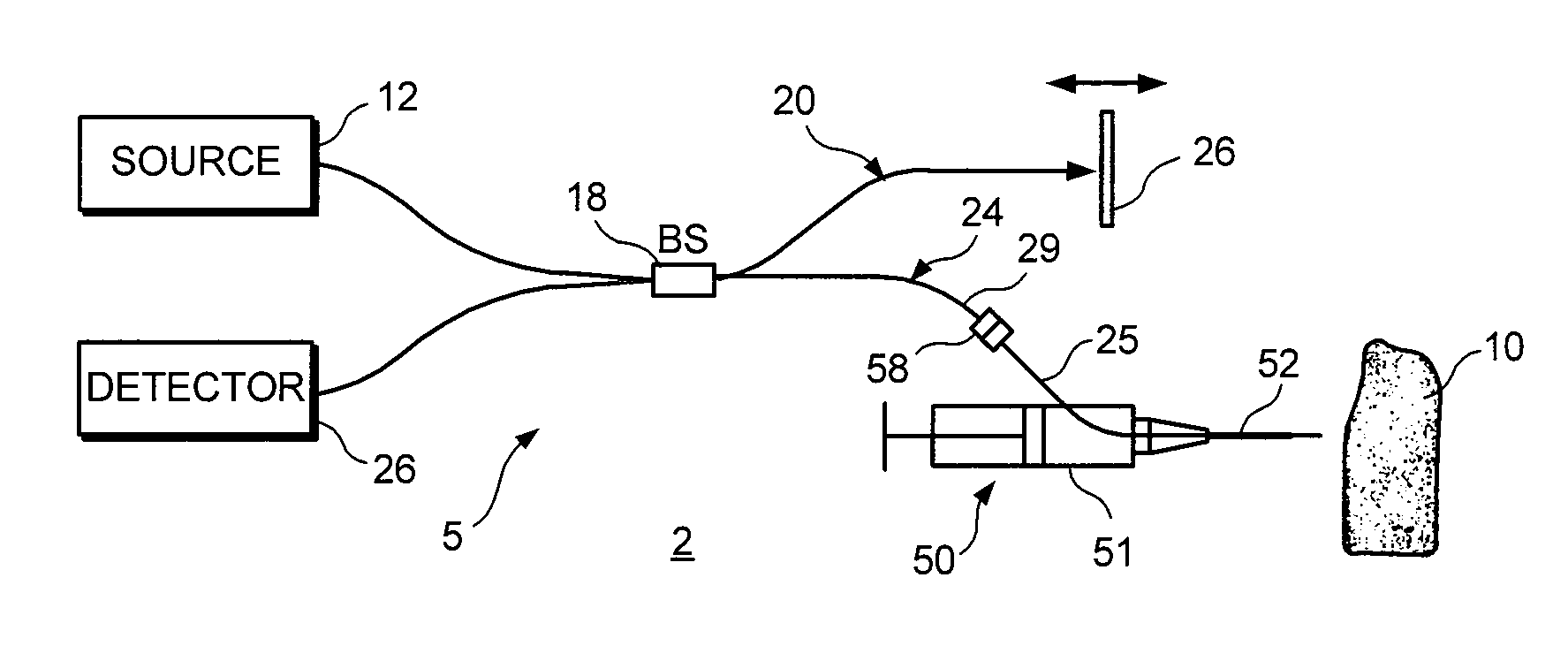
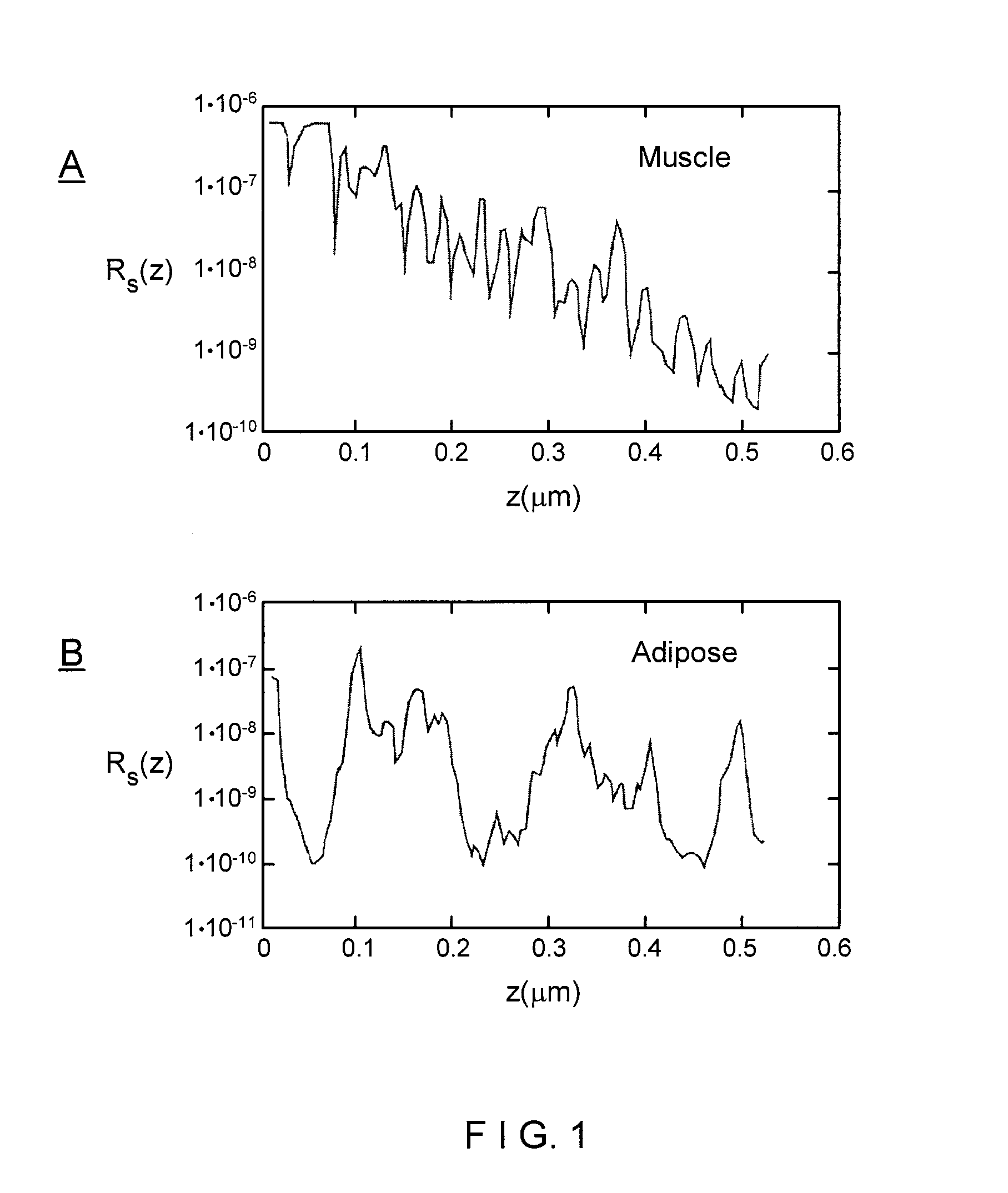
[0047]In accordance with the system of the present invention, FIG. 2 illustrates an tissue identification system 2 according to one embodiment of the present invention for tissue 10 identification using interferometric ranging. The tissue identification system 2 utilizes a one-dimensional data set in order to identify tissue. Unlike many prior art systems, which use two-dimensional data in order to acquire sufficient information to identify tissue, the tissue identification system 2 is able to identify tissue using a one-dimensional data set. Differences between two types of tissue may be understood from a one-dimensional data set. For example, FIG. 1 illustrates two graphs that represent a one-dimensional interferometric ranging axial scan of two different tissue types. As can be seen from these graphs, adipose tissue (shown in the bottom graph) has a significantly different axial reflectance profile as compared to the axial reflectance profile of muscle tissue (shown in the top gr...


PUM


Abstract

An apparatus for needle biopsy with real time tissue differentiation using one dimensional interferometric ranging imaging, comprising a biopsy device having a barrel and a needle, an optical fiber inserted in the needle, and a fiber optic imaging system connected to the optical fiber. The imaging system obtains images and compares the optical properties and patterns to a database of normalized tissue sample images to determine different tissue types. The physician performing the biopsy obtains feedback via a feedback unit associated with the biopsy device and which is connected to the imaging system. The feedback unit can provide visual, audible or vibratory feedback as to tissue type encountered when the needle is inserted toward the target tissue. The feedback unit can be programmed for different biopsy procedures so that the user can actuate a button to select a display or other feedback mechanism for the desired procedure and anticipated tissue to be encountered.

Description

RELATED APPLICATIONS[0001]This application claims priority to U.S. Provisional Application Ser. No. 60 / 442,392 filed Jan. 24, 2003, entitled “Devices and Methods for Tissue Identification Using Low-Coherence Interferometry,” which is incorporated by reference herein in its entirety.BACKGROUND OF THE INVENTION[0002]1. Field of the Invention[0003]The present invention relates to an apparatus and method for identifying tissue types using interferometric ranging during needle biopsy. More particularly, the present invention relates to an imaging system including a needle probe and algorithms for detecting various tissue types during a biopsy. Also provided is a method for differentiating tissue types using the imaging system.[0004]2. Background of the Invention[0005]A significant cause of inefficiency of intraoperative and biopsy procedures is the inability of a physician to identify tissue type by gross inspection. For example, head and neck surgeries, the inability to differentiate mu...

Claims


Application Information

Patent Timeline
Patent Type & Authority Patents(United States)
IPC IPC(8): A61B5/00A61BA61B6/00
CPCA61B5/0066A61B5/415A61B5/6852A61B5/418A61B5/6848A61B5/416
Inventor TEARNEY, GUILLERMO J.SHISHKOV, MILEN STEFANOVIFTIMIA, NICUSOR V.BOUMA, BRETT E.PITMAN, MARTHA
Owner THE GENERAL HOSPITAL CORP
Why Eureka
Try Eureka