Method for establishing animal model of tracheostenosis and equipment thereof

An animal model, tracheal stenosis technology, applied in the medical field, can solve the problems of complicated steps, damage to the tracheal wall, poor repeatability, etc., and achieve the effect of good repeatability and easy operation

Inactive Publication Date: 2013-04-03
THE FIRST AFFILIATED HOSPITAL OF GUANGZHOU MEDICAL UNIV (GUANGZHOU RESPIRATORY CENT) +2
View PDF5 Cites 2 Cited by
  • Summary
  • Abstract
  • Description
  • Claims
  • Application Information

AI Technical Summary

Problems solved by technology

In recent years, various methods have been tried to establish animal models of tracheal stenosis at home and abroad, mainly including thermal ablation to damage the tracheal wall, tracheotomy and suture, mechanical and chemical damage methods, etc. Generall

Method used


Image

  • Method for establishing animal model of tracheostenosis and equipment thereof
  • Method for establishing animal model of tracheostenosis and equipment thereof
  • Method for establishing animal model of tracheostenosis and equipment thereof

Examples


Embodiment Construction

[0026] The present invention will be described in further detail below in conjunction with the accompanying drawings and specific embodiments.

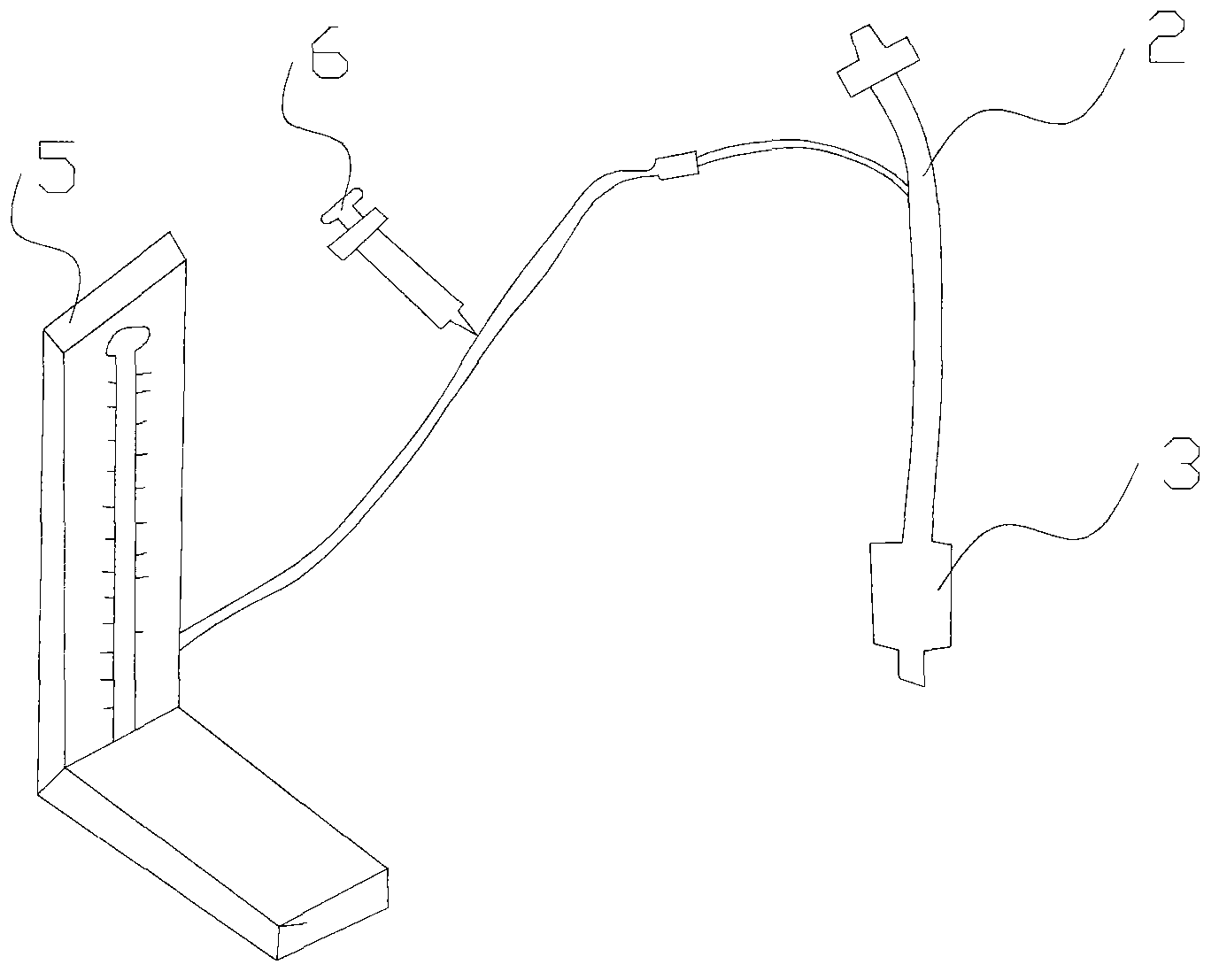
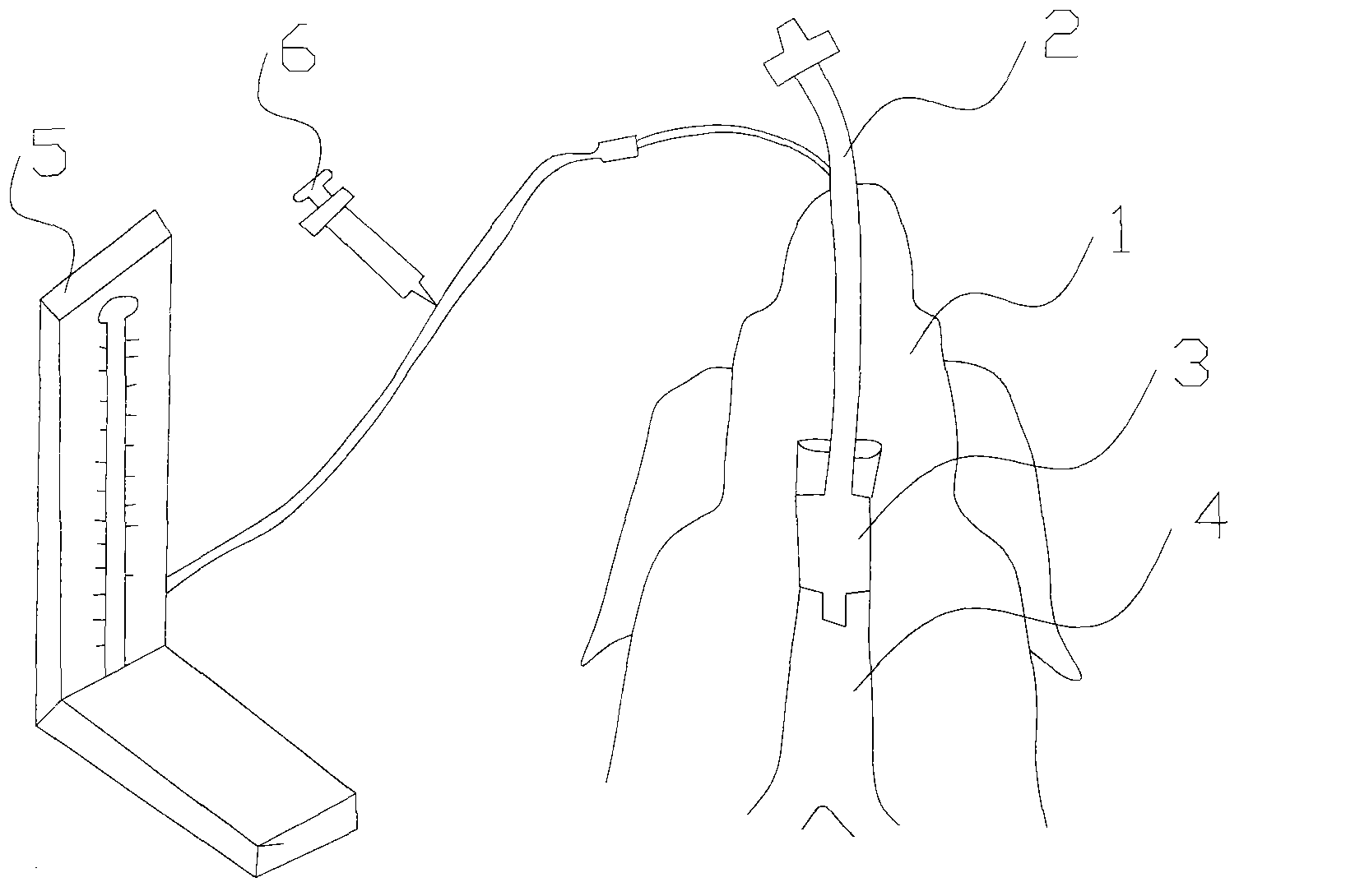
[0027] refer to figure 1 As shown, the present invention provides a device for establishing an animal model of tracheal stenosis, which includes an intubation catheter 2, an inflatable balloon 3, a manometer 5 and a syringe 6, and the inflatable balloon 3 is installed on the intubation catheter 2 At one end, the manometer 5 and the syringe 6 are installed at the other end of the intubation catheter 2, wherein the syringe 6 is used to inflate the inflatable balloon, and the manometer 5 is used to measure the pressure of the inflatable balloon The shape of the inflatable balloon 3 after inflation is cylindrical; the manometer 5 is a mercury sphygmomanometer;

[0028] refer to Figure 5 Shown, on the whole, the process of establishing tracheal stenosis animal model of the present invention is:

[0029] First choice, choose an intubati...


PUM


Abstract

The invention discloses a method for establishing an animal model of tracheostenosis and equipment thereof. The method comprises the steps of: selecting one intubation catheter with a sacculus; carrying out orotracheal intubation on an experimental animal, injecting gas in the sacculus and fixing the intubation catheter, wherein the air pressure injected into the sacculus is 175-225 mmHg, and the intubation catheter is kept for 18-25h; and pulling out the intubation catheter, wherein 1-3 weeks after the intubation catheter is pulled out, tracheostenosis is formed because of local granulation scar tissue proliferation and the tracheal collapse of the tracheal wall of the experimental animal. The invention also discloses the equipment for realizing the method. The method and the equipment, disclosed by the invention, have the advantages that simplicity and convenience are brought in operation, the repeatability is good and the real process of the generation of tracheostenosis of a clinical patient after endotracheal intubation is simulated, therefore the stable and reliable animal model of tracheostenosis for the research of benign airway stenosis is provided.

Description

technical field [0001] The invention relates to the field of medical technology, in particular to a method and equipment for establishing an animal model of tracheal stenosis through tracheal intubation. Background technique [0002] Tracheal stenosis is a common complication after long-term or inappropriate tracheal intubation, and post-intubation tracheal stenosis is also one of the most common types of benign airway stenosis. The occurrence of tracheal stenosis after intubation is mainly related to factors such as too high pressure of the air bag of the tracheal tube, too long intubation time, and improper intubation operation. Prolonged pressure on the tracheal wall leads to mucosal edema and necrosis, ulcer formation, and even chondritis or cartilage necrosis, which eventually leads to local granulation scar tissue hyperplasia and / or tracheal collapse to form tracheal stenosis. Despite the widespread use of low-pressure, high-volume cuffed endotracheal tubes, post-intu...

Claims


Application Information

Patent Timeline
IPC IPC(8): A61M16/04
Inventor 李时悦苏柱泉陈小波
Owner THE FIRST AFFILIATED HOSPITAL OF GUANGZHOU MEDICAL UNIV (GUANGZHOU RESPIRATORY CENT)
Why Eureka
Try Eureka