Fluorescence angiography fundus image extraction method based on NGC-ACM

A fundus image, extraction method technology, applied in the field of image recognition, to achieve the effect of improving the effect and quality, and good global segmentation effect

Active Publication Date: 2015-01-21
CENT SOUTH UNIV
View PDF1 Cites 12 Cited by
  • Summary
  • Abstract
  • Description
  • Claims
  • Application Information

AI Technical Summary

Problems solved by technology

However, most existing algorithms only segment images based on either region or boundary information, and using this incomple

Method used


Image

  • Fluorescence angiography fundus image extraction method based on NGC-ACM
  • Fluorescence angiography fundus image extraction method based on NGC-ACM
  • Fluorescence angiography fundus image extraction method based on NGC-ACM

Examples


Embodiment Construction

[0062] The present invention will be further described below in conjunction with the accompanying drawings and embodiments.

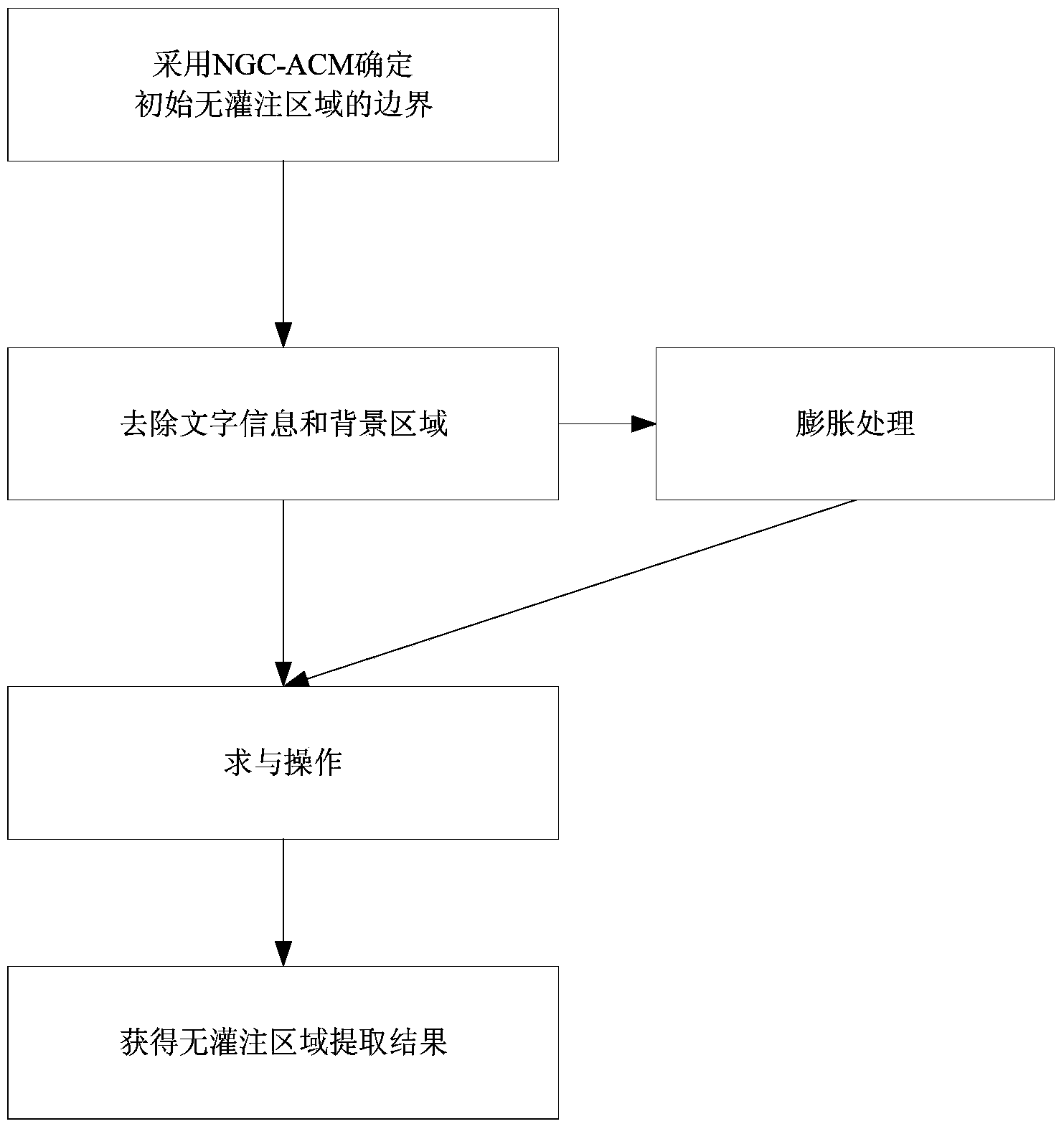
[0063] Such as figure 1 Shown is a schematic flow chart of the method of the present invention, a method for extracting fundus images based on NGC-ACM fluorescein contrast, comprising the following steps:

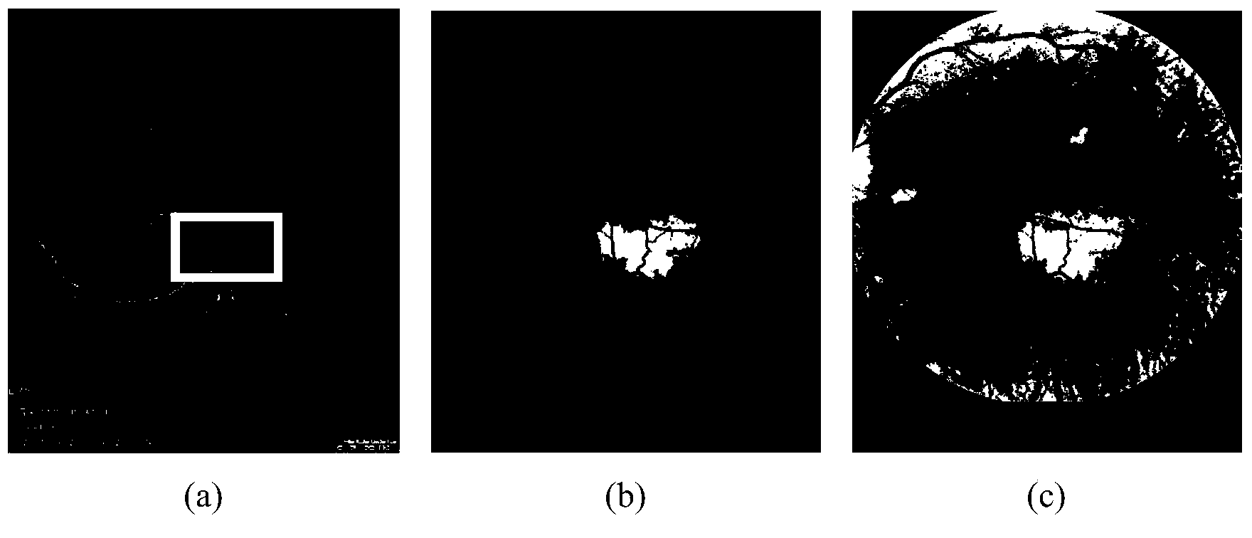
[0064] Step 1: Manually select the initial non-perfusion area in the fluorescein contrast-enhanced fundus image, and use the NGC-ACM algorithm to determine the boundary of the initial non-perfusion area; the results are as follows figure 2 As shown, where (a) is the initial non-perfusion area manually selected, (b) is the local extraction result obtained only near the initialization area, and the effect is good, (c) is running NGC- The global result obtained by the ACM algorithm is poor, and a large number of invalid extraction areas appear.

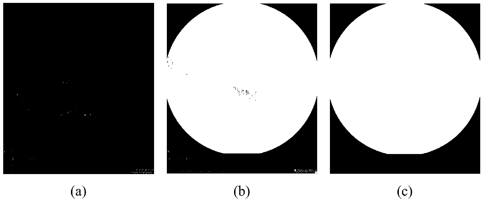
[0065] Step 2: remove the text information and the background area in the area included in the boundary o...


PUM


Abstract

The invention discloses a fluorescence angiography fundus image extraction method based on NGC-ACM. The method includes the following steps that firstly, a fluorescence angiography fundus image is processed, a region of interest is selected by a user, and a boundary of a non-filling region is preliminarily determined; then, through fuzzy division of the fluorescence angiography fundus image, the range in which the non-filling region surely exists is extracted; then, a binarized template is manufactured for the fluorescence angiography fundus image, and boundary information in a region extraction result is eliminated; finally, through mathematical morphological processing of the extracted boundary of the non-filling region and the extracted range in which the non-filling region exists, an extraction result of the non-filling region is acquired finally. The overall extraction effect and quality of the non-filling region are greatly improved.

Description

technical field [0001] The invention belongs to the technical field of image recognition, and relates to medical image processing, in particular to an NGC-ACM-based method for extracting fundus images of fluorescein contrast. Background technique [0002] In today's society, the number of people with diabetes is increasing with the improvement of living standards, resulting in a very high incidence of diabetic retinopathy (diabetic retinopathy, DR, referred to as "sugar net"). At the same time, the fundus retinal imaging technology developed in recent years provides a simple and non-invasive fundus detection method for diabetic reticulum patients, which is easily accepted by patients and has become an important basis for doctors to diagnose ophthalmic diseases. However, the current diagnosis methods in hospitals mainly observe and analyze the fluorescein contrast fundus images with the naked eyes of doctors, which requires high experience and experience of doctors, and the p...

Claims


Application Information

Patent Timeline
IPC IPC(8): G06T7/00G06T5/00
Inventor 向遥李暄邱从贤朱承璋邹北骥陈慧慧高玲
Owner CENT SOUTH UNIV
Why Eureka
Try Eureka