Direct three-dimensional scanning method and system for physiological motion boundary of oral soft tissue

A three-dimensional scanning and moving boundary technology, applied in the field of intraoral scanning, can solve the problems of low success rate, low efficiency, inaccurate impression of oral soft tissue functional boundary, etc., and achieve the effect of fast scanning speed, simple operation and high precision

Pending Publication Date: 2020-08-25
SICHUAN UNIV
View PDF6 Cites 3 Cited by
  • Summary
  • Abstract
  • Description
  • Claims
  • Application Information

AI Technical Summary

Problems solved by technology

However, although the existing intraoral scanning methods can scan the soft tissues in the oral cavity, the main problems are: first, the three-dimensional intraoral scanner cannot obtain the functional boundary of the soft tissues; The impression is inaccurate; the third is that the efficiency and success rate of making an impression of the functional boundary of ...

Method used


Image

  • Direct three-dimensional scanning method and system for physiological motion boundary of oral soft tissue
  • Direct three-dimensional scanning method and system for physiological motion boundary of oral soft tissue
  • Direct three-dimensional scanning method and system for physiological motion boundary of oral soft tissue

Examples


Embodiment 1

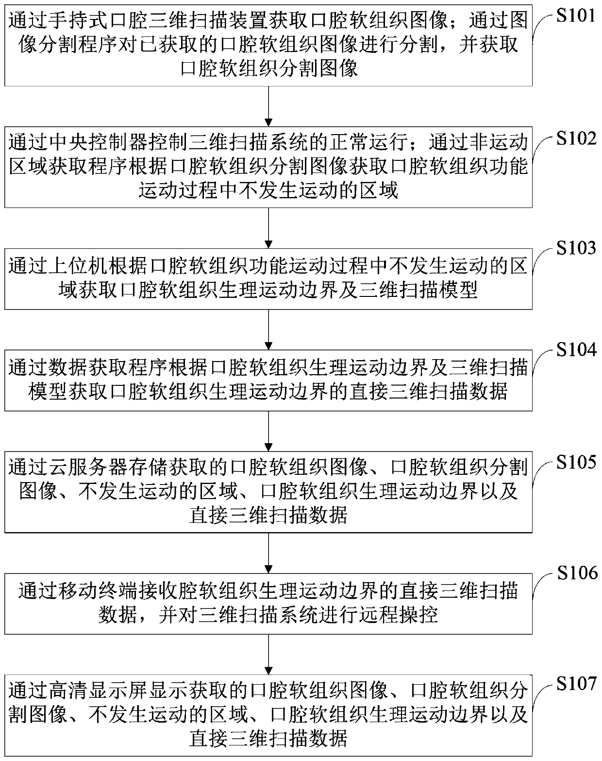
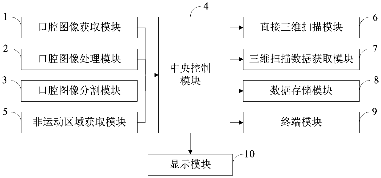
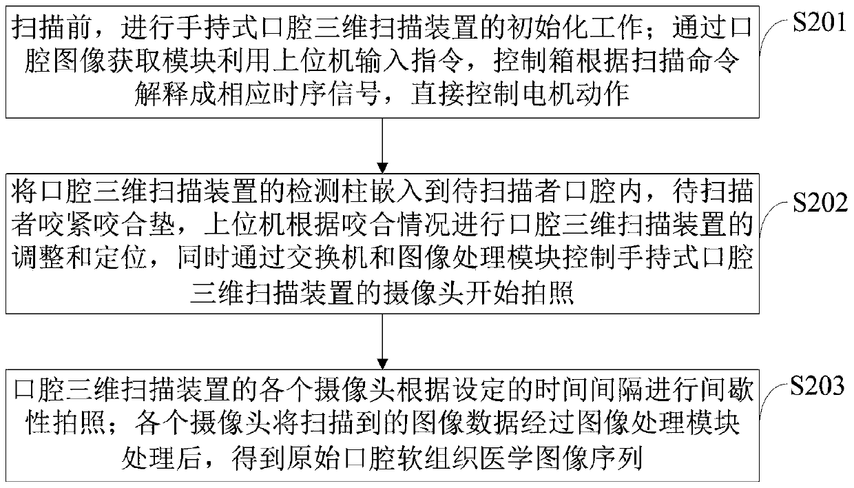
[0085] The direct three-dimensional scanning method of the oral soft tissue physiological motion boundary provided by the embodiment of the present invention is as follows: figure 1 As shown, as a preferred embodiment, such as image 3 As shown, the method for acquiring soft tissue images of the oral cavity through a handheld oral three-dimensional scanning device provided by the embodiment of the present invention includes:

[0086] S201. Before scanning, initialize the hand-held oral three-dimensional scanning device; use the upper computer to input commands through the oral image acquisition module, and the control box interprets the scanning commands into corresponding timing signals to directly control the motor action.

[0087] S202, embed the detection column of the oral three-dimensional scanning device into the oral cavity of the person to be scanned, and the person to be scanned bites the occlusal pad tightly, and the host computer adjusts and positions the oral thre...

Embodiment 2

[0094] The direct three-dimensional scanning method of the oral soft tissue physiological motion boundary provided by the embodiment of the present invention is as follows: figure 1 As shown, as a preferred embodiment, such as Figure 4 As shown, the method provided by the embodiment of the present invention to segment the acquired oral soft tissue image through the image segmentation program and obtain the segmented oral soft tissue image includes:

[0095] S301, acquiring the dental medical image sequence I through an image segmentation program i , and intercept the effective range of pixel values.

[0096] S302, use the active contour method to semi-automatically segment the dental image, and use it as the data I for the training of the generative confrontation network i '.

[0097] S303, raw stomatology image I i Stomatological image obtained by segmentation with active contour method I i 'Send it into the confrontational generative network for training and learning, ...


PUM


Abstract

The invention belongs to the technical field of oral scanning, and discloses a direct three-dimensional scanning method and system for a physiological motion boundary of oral soft tissue. The direct three-dimensional scanning system for the physiological motion boundary of the oral soft tissue comprises an oral image acquisition module, an oral image processing module, an oral image segmentation module, a central control module, a non-motion area acquisition module, a direct three-dimensional scanning module, a three-dimensional scanning data acquisition module, a data storage module, a terminal module and a display module. The scanning system provided by the invention is high in scanning speed, high in precision and simple to operate; an active contour method is used for semi-automatically carrying out labeling segmentation on an oral medical image serving as a training set, and the semi-automatic method improves the accuracy and timeliness of labeling segmentation; and a confrontation generation network is used for automatically generating a segmentation result of the oral medical image, so that the complete and effective expression of the healthy oral physiological structure isbalanced in key indexes such as segmentation precision, efficiency, stability and robustness.

Description

technical field [0001] The invention belongs to the technical field of intraoral scanning, and in particular relates to a direct three-dimensional scanning method and system for the physiological movement boundary of oral soft tissue. Background technique [0002] Typically, dental treatment is performed by an impression procedure in which a plaster model of a patient's teeth is taken in a dental office or similar. However, this impression process may be accompanied by various problems such as consumption of impression material, cross-contamination, breakage of the assembled model, etc. Therefore, the two-dimensional pattern of teeth is formed by radiating radiation, which has the disadvantages of exposing the patient to a large amount of radiation and poor precision. Then, three-dimensional dental scanners were developed and widely used. However, although the existing intraoral scanning methods can scan the soft tissues in the oral cavity, the main problems are: first, th...

Claims


Application Information

Patent Timeline
IPC IPC(8): A61B5/00
CPCA61B5/0088A61B5/0033
Inventor 肖诗梦张曜耀张雨薇林意澄丁一
Owner SICHUAN UNIV
Why Eureka
Try Eureka