Method of image quality assessment to produce standardized imaging data

a technology of image quality assessment and imaging data, applied in the field of medical imaging, can solve the problems of limited cervical imagery quality, incorrect instrument positioning, and incorrect instrument settings

Inactive Publication Date: 2008-09-18
CADES SCHUTTE A LIMITED LIABILITY LAW PARTNERSHIP
View PDF8 Cites 106 Cited by
  • Summary
  • Abstract
  • Description
  • Claims
  • Application Information

AI Technical Summary

Problems solved by technology

The limited quality of cervical imagery can be attributed to several factors, including: incorrect instrument settings, incorrect instrument positioning, glint, blur due to poor focus, and physical contaminants.
Glint (specular reflection) eliminates the color information in affected pixels and can therefore introduce artifacts in feature extraction algorithms.
Instrument settings that result in an inadequate dynamic range (defined below) or overly constrained (too small) region of interest can reduce or eliminate pixel information and thus make image analysis algorithms unreliable.
Poor focus causes image blur with a consequent loss of texture information.
In addition, a variety of physical contaminants, such as blood, can obscure the desired scene, and reduce or eliminate diagnostic information from affected areas.

Method used


Image

  • Method of image quality assessment to produce standardized imaging data
  • Method of image quality assessment to produce standardized imaging data
  • Method of image quality assessment to produce standardized imaging data

Examples


Embodiment Construction
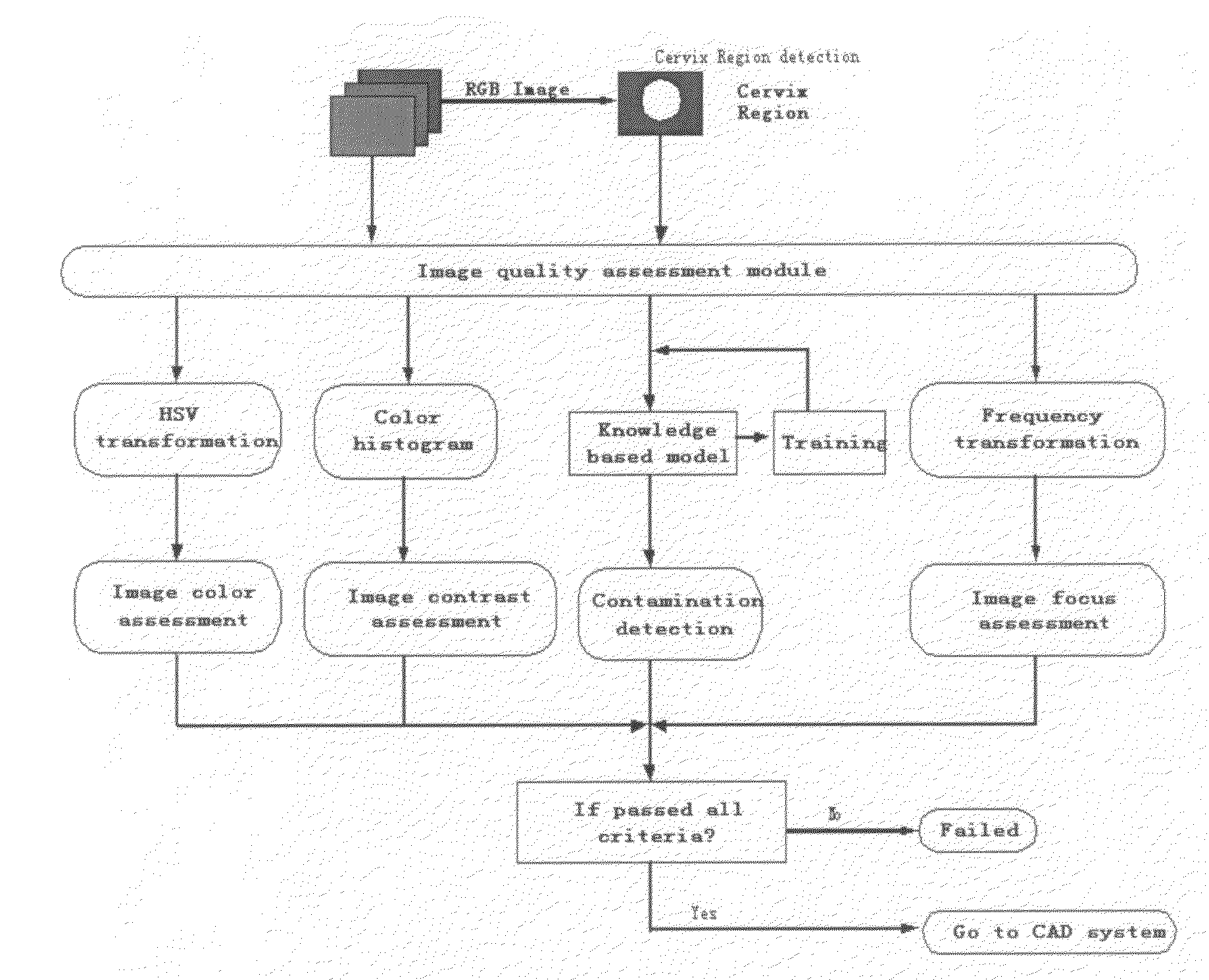
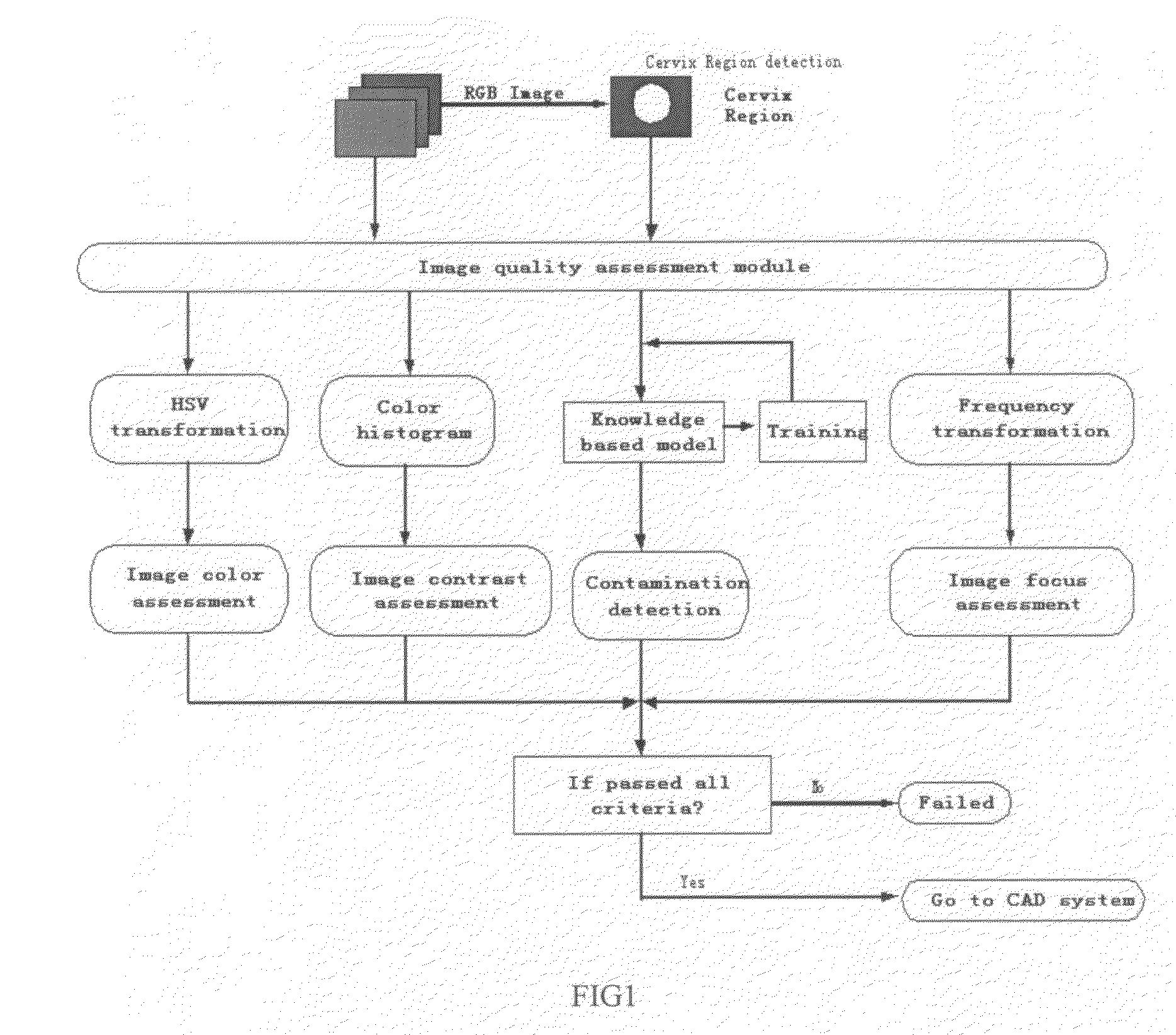
1. System Framework

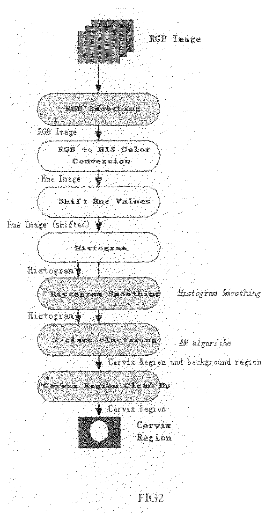
[0034]The presently preferred embodiment of the invention described herein preferably starts from an RGB (Red-Green-Blue) color space image from a digital colposcope. The input image is a glare free RGB image of a uterine cervix. Glare free imagery can be obtained either by cross-polarized (XP) image acquisition or glare removal pre-processing (Lange H.; Automatic glare removal in reflectance imagery of the uterine cervix; SPIE Medical Imaging 2005; SPIE Proc. 5747, 2005, incorporated herein by reference).

[0035]The invention preferably comprises a framework of robust, real-time, industry-oriented algorithms to carry out the invention using statistical, morphological and signal processing methods. First, a Region-Of-Interest (ROI), preferably the cervix, is detected using a hue color cluster that discriminates between cervix and background. Then an adaptive peak-removing histogram equalization algorithm is used to assess the contrast. Following the contrast assessm...


PUM


Abstract

Automated image quality assessment methods, which include locating a region of interest, region assessment, contrast assessment, blur assessment, and contaminant detection, on video data and high-resolution imagery. Where the blur assessment is performed without a reference image by dividing the region into non-overlapping block, measuring the wavenumber frequency of the blocks and calculating the ratio of the low frequency to high frequency areas.

Description

[0001]This application claims priority to U.S. provisional patent application 60 / 918,527 filed on Mar. 16, 2007.TECHNICAL FIELD[0002]This invention generally relates to medical imaging and more specifically to image processing to achieve high-quality standardized digital imagery for use in archive-quality medical records and Computer-Aided-Diagnosis (CAD) systems.BACKGROUND ART[0003]Although this invention is being disclosed in connection with cervical cancer, it is applicable to many other areas of medicine. Uterine cervical cancer is the second most common cancer in women worldwide, with nearly 500,000 new cases and over 270,000 deaths annually (http: / / www-depdb.iarc.fr / globocan2002.htm, incorporated herein by reference) Colposcopy is a diagnostic method used to detect cancer precursors and cancer of the uterine cervix (B. S. Apgar, Brotzman, G. L. and Spitzer, M., Colposcopy: Principles and Practice, W. B. Saunders Company: Philadelphia, 2002, incorporated herein by reference). C...

Claims


Application Information

Patent Timeline
Patent Type & Authority Applications(United States)
IPC IPC(8): G06K9/00G06K9/40G06V10/56
CPCA61B5/0084G06T5/007G06T5/40G06T5/50G06T7/0002G06K9/4652G06T2207/10152G06T2207/30004G06T2207/30168G06T2207/10024G06T2207/10016G06V10/56
Inventor GU, JIALI, WENJINGHARGROVE, JOHNWOLTERS, ROLF HOLGER
Owner CADES SCHUTTE A LIMITED LIABILITY LAW PARTNERSHIP
Why Eureka
Try Eureka