Medical devices and methods of placement

a technology of medical devices and methods, applied in the field of medical devices, can solve the problems of increasing the complexity of the task, increasing the risk of the task, and requiring significant skill for the patient's trachea

Active Publication Date: 2017-07-27
WM & DG
View PDF0 Cites 34 Cited by
  • Summary
  • Abstract
  • Description
  • Claims
  • Application Information

AI Technical Summary

Problems solved by technology

Because human anatomy varies significantly from a patient to a patient, properly placing a medical device in a patient's trachea requires a significant skill and is a task laced with inherent risk.
The task becomes even more complicated because the insertion procedure may have to be performed immediately at an accident site, on pediatric patients, in a nursing home, on a battlefield or at a natural disaster site where many patients have to be attended at the same time.
These devices provide accuracy for initial placement, but do not provide continuous visualization or mobility of the image after a medical device has been placed in a patient.
Newer devices, such as Vivasight SL or DL endotracheal tubes, provide continuous visualization, but are costly because they depend on a single use of disposable cameras and they are not transferrable from one medical device to another.

Method used


Image

  • Medical devices and methods of placement
  • Medical devices and methods of placement
  • Medical devices and methods of placement

Examples


Embodiment Construction

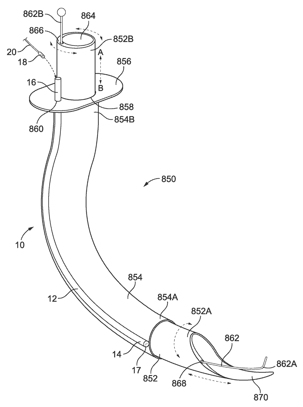
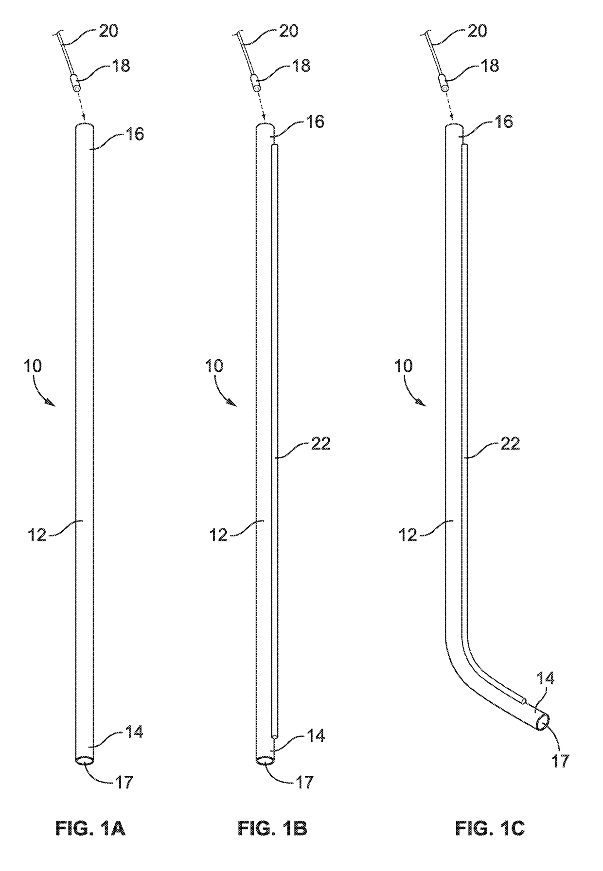
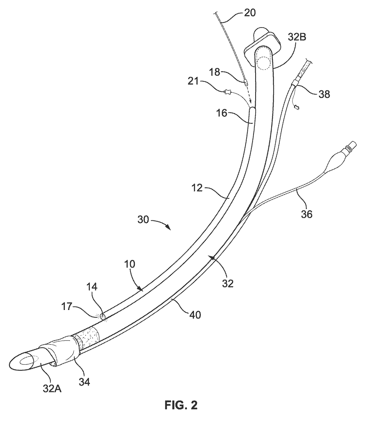
[0097]The present invention provides improved medical devices equipped with a visualization device for intubation, ventilation, feeding and monitoring of a patient. The present invention also provides methods for rapid and accurate placement of a medical device in a patient and remote continuous real-time monitoring of the patient after the placement.

[0098]These medical devices are equipped with a visualization device in which a camera is placed in a separate sealed camera tube. As the camera does not come in contact with a patient, there is no need to sterilize the camera and the same camera can be reused in many applications. Thus, the same camera can be switched between different medical devices which monitor internal organs such as medical devices that are placed in patient's airway, larynx, gastrointestinal tract, chest or vaginal cavity. In some embodiments, the camera is disposable.

[0099]One embodiment provides a visualization device as shown in FIG. 1A and its further embodi...


PUM


Abstract

The present invention provides medical devices comprising at least one visualization device sealed to, attached to or otherwise combined with at least one of the following second devices: an oral airway, ventilating mask, urinary catheter, trocar, a tool tube, and a medical glove. The present invention also provides methods for rapid and accurate placement of a medical device in a patient and continuous real time monitoring, including a remote monitoring, of the patient after the placement.

Description

CROSS-REFERENCE TO RELATED APPLICATIONS[0001]This application is a continuation-in-part of U.S. patent application Ser. No. 15 / 163,361 filed on May 24, 2016, which is a continuation-in part of U.S. patent application Ser. No. 14 / 798,751 filed on Jul. 14, 2015, which is a continuation-in-part of U.S. patent application Ser. No. 14 / 455,470 filed Aug. 8, 2014, the entire disclosures of which are incorporated herein.FIELD OF THE INVENTION[0002]The invention provides various medical devices, each with a camera placed in a camera tube, which allows for a camera to provide continuous visualization for each device during and after placement in a patient. A sound device, such as a microphone, is incorporated in many devices and provides continuous monitoring of breath and heart sounds in a patient. The camera and microphone do not contact the patient's tissues and thus, do not require sterilization. The continuous visualization and sound monitoring of the patient are in real time and enable ...

Claims


Application Information

Patent Timeline
Patent Type & Authority Applications(United States)
IPC IPC(8): A61B1/00A61B1/04
CPCA61B1/00A61B1/00163A61B1/04A61B1/00112A61B1/2676A61B1/00128A61B1/307A61B1/267A61B1/2673A61B1/00121A61B1/00016A61B1/00078A61B1/00135A61B1/00137A61B1/0014A61B1/00142A61B1/00154A61B1/015A61B1/018A61B1/05A61B1/07A61B1/233A61B1/303A61M16/0434A61M16/0461A61M16/0463A61M16/0465A61M16/0488A61M16/0497A61M16/0666A61M16/0409A61M16/0456A61M16/0459
Inventor MOLNAR, ROBERT
Owner WM & DG
Why Eureka
Try Eureka