Vacuum syringe assisted biopsy device

a technology of syringe and biopsy device, which is applied in the field of biopsy devices, can solve the problems of high patient cost and high level of trauma, open biopsy carries a relatively higher risk of infection and bleeding, and is difficult to read future mammograms

Active Publication Date: 2010-11-09
DEVICOR MEDICAL PROD
View PDF63 Cites 77 Cited by
  • Summary
  • Abstract
  • Description
  • Claims
  • Application Information

AI Technical Summary

Benefits of technology

[0012]The present invention addresses these and other problems of the prior art by providing a biopsy device that has a probe cannula that is inserted into tissue to obtain a core biopsy sample by translating a cutter with the probe cannula. Vacuum assistance to prolapse tissue for sampling is advantageously provided by an integral vacuum container whose internal pressure is reduced from atmospheric pressure by actuation of a single motor that also translates the cutter to sever biopsy samples.

Problems solved by technology

An open biopsy is a surgical procedure that is usually done as an outpatient procedure in a hospital or a surgical center, involving both high cost and a high level of trauma to the patient.
Open biopsy carries a relatively higher risk of infection and bleeding than does percutaneous biopsy, and the disfigurement that sometimes results from an open biopsy may make it difficult to read future mammograms.
Further, the aesthetic considerations of the patient make open biopsy even less appealing due to the risk of disfigurement.
Given that a high percentage of biopsies show that the suspicious tissue mass is not cancerous, the downsides of the open biopsy procedure render this method inappropriate in many cases.
However, a shortcoming of FNA is that only a small number of cells are obtained through the procedure, rendering it relatively less useful in analyzing the suspicious tissue and making an assessment of the progression of the cancer less simple if the sample is found to be malignant.
With a long probe, the rate of sample taking is limited not only by the time required to rotate or reposition the probe but also by the time needed to translate the cutter.
Such tethers may tend to impede positioning of the biopsy device, introduce tripping hazards, and increase set up time.

Method used


Image

  • Vacuum syringe assisted biopsy device
  • Vacuum syringe assisted biopsy device
  • Vacuum syringe assisted biopsy device

Examples


Embodiment Construction

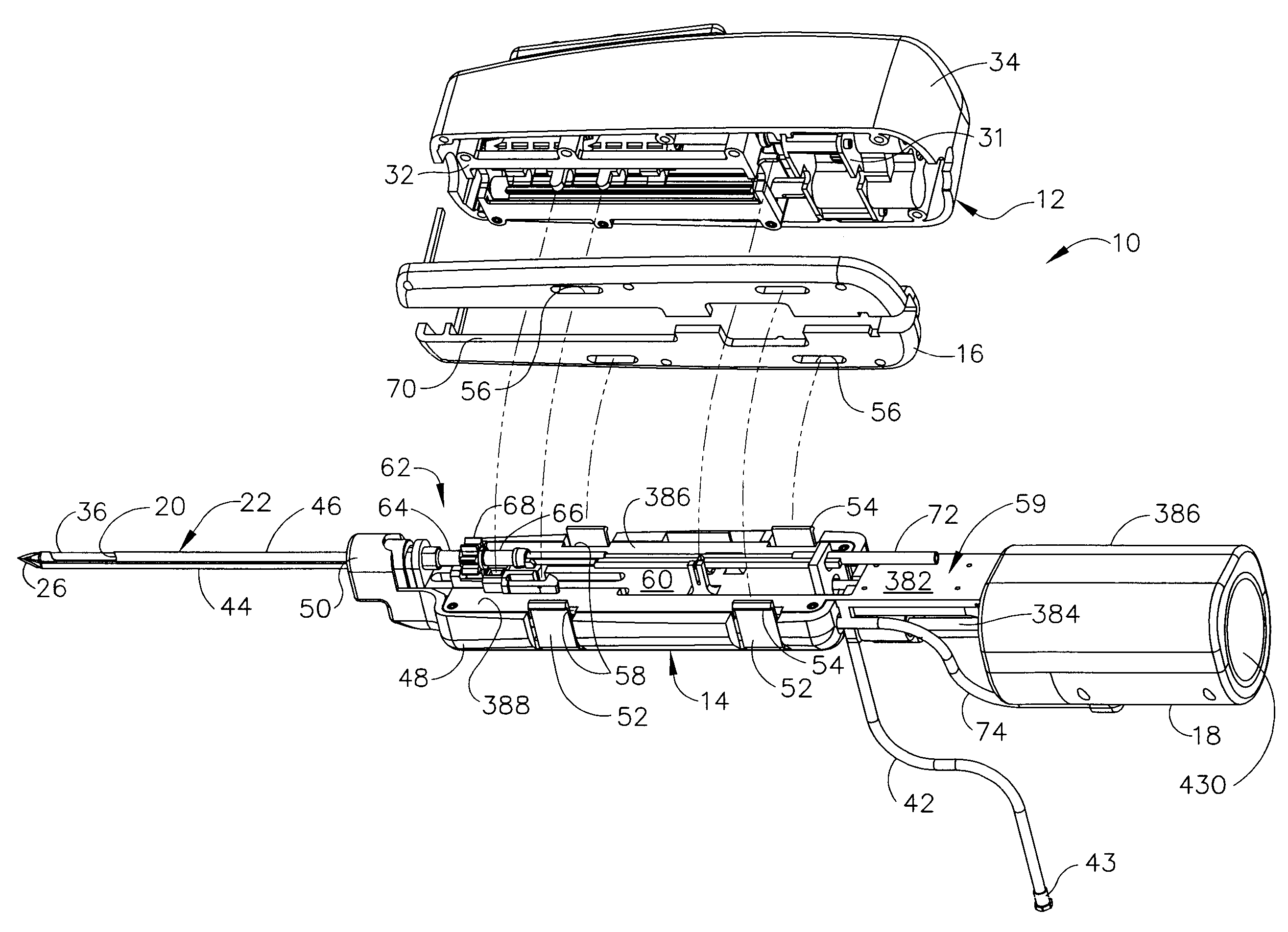
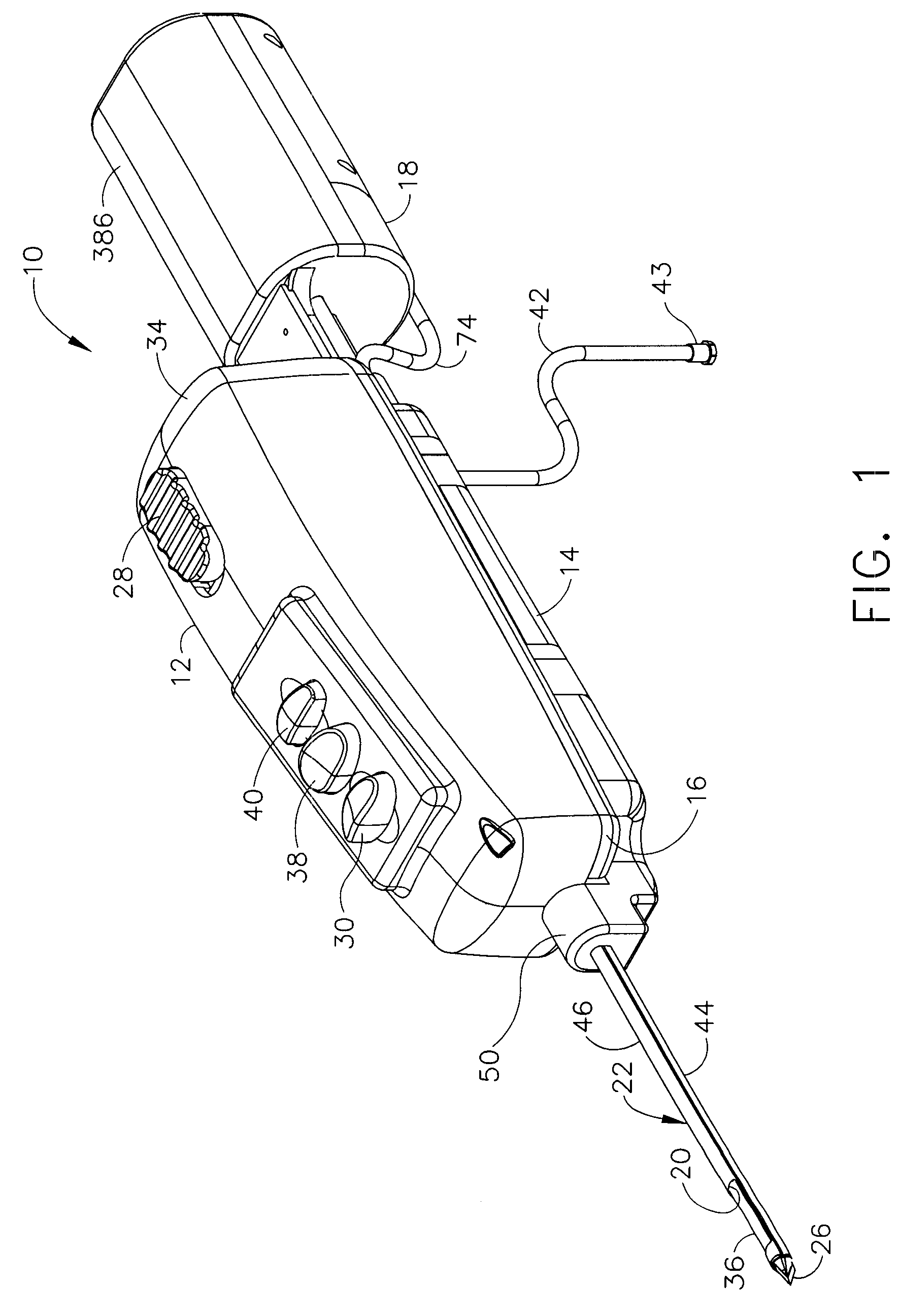
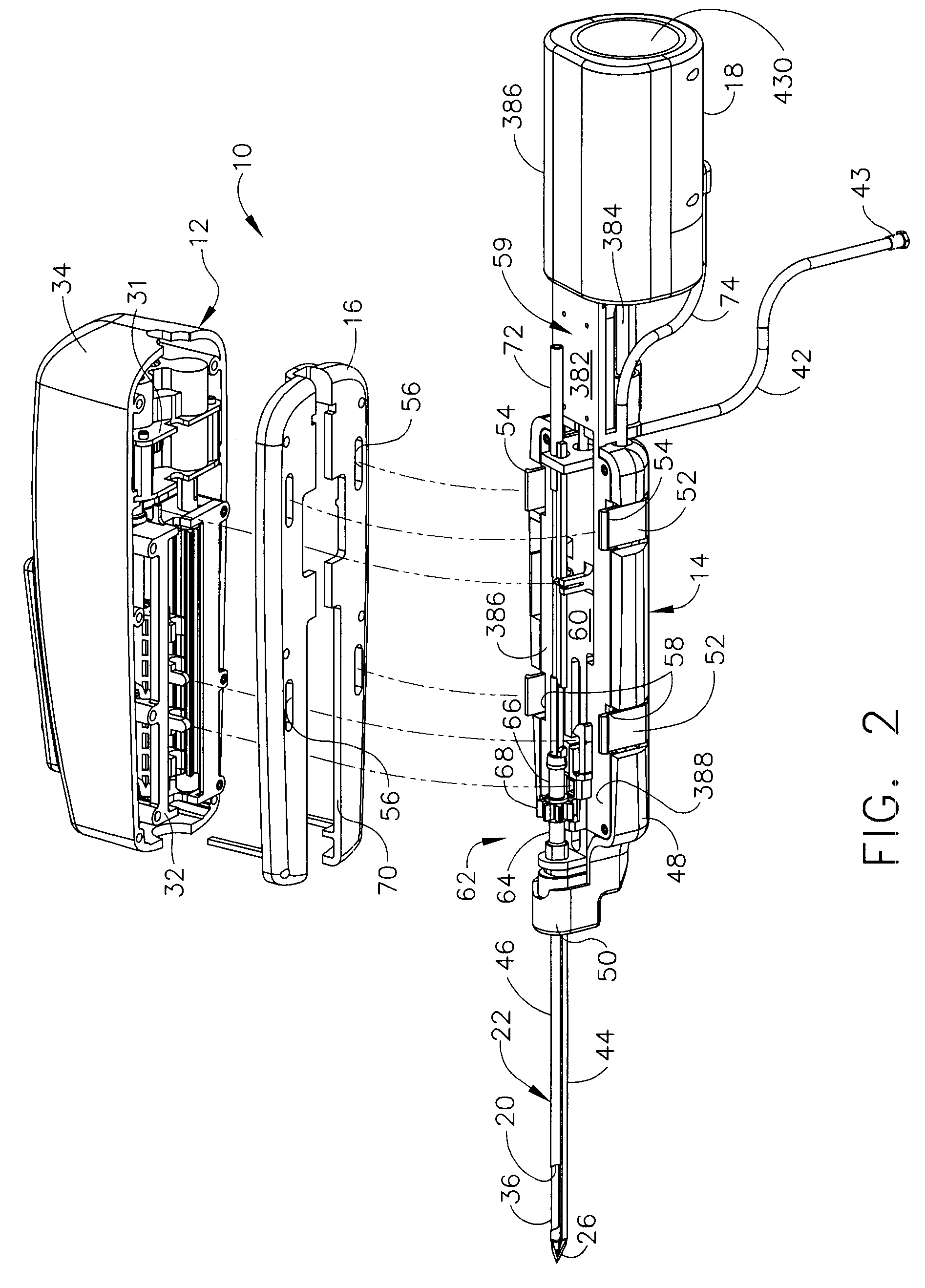
[0038]Turning to the Drawings, wherein like numerals denote like components throughout the several views, in FIGS. 1-3, a biopsy device 10 includes a reusable handpiece 12, and a disposable probe assembly 14. A lower handle tray 16 is disassembled from upper portions of the reusable handpiece 12 to expose portions that operably engage the disposable probe assembly 14. A vacuum syringe assembly 18 is a proximal portion of the disposable probe assembly 14 that is also actuated by the reusable handpiece 12. With the close proximity of the source of vacuum, the amount of vacuum line that needs to be evacuated is minimized, enabling a modestly sized vacuum syringe assembly 18 to effect vacuum assistance to prolapse tissue into a side aperture 20 of a probe cannula 22 of the disposable probe assembly 14. In FIG. 3, further economy is realized by employing one DC motor 24 in the reusable handpiece 12 to accomplish the severing of tissue samples as well as actuating the vacuum syringe assem...


PUM


Abstract

A biopsy device and method are provided for obtaining a tissue sample, such as a breast tissue biopsy sample. The biopsy device includes a disposable probe assembly with an outer cannula having a distal piercing tip, a cutter lumen, and a cutter tube that rotates and translates past a side aperture in the outer cannula to sever a tissue sample. The biopsy device also includes a reusable handpiece with an integral motor and power source to make a convenient, untethered control for use with ultrasonic imaging. The reusable handpiece incorporates a probe oscillation mode to assist when inserting the distal piercing tip into tissue. The motor also actuates a vacuum syringe in coordination with movement of the cutter tube to provide vacuum assistance in prolapsing tissue and retracting tissue samples.

Description

CROSS REFERENCE TO RELATED APPLICATIONS[0001]The present application is a continuation-in-part of the co-pending and commonly-owned U.S. patent application Ser. No. 11 / 198,558, “BIOPSY DEVICE WITH REPLACEABLE PROBE AND INCORPORATING VIBRATION INSERTION ASSIST AND STATIC VACUUM SOURCE SAMPLE STACKING RETRIEVAL” to Hibner et al., filed 5 Aug. 2005, the disclosure of which is hereby incorporated by reference in its entirety.FIELD OF THE INVENTION[0002]The present invention relates in general to biopsy devices, and more particularly to biopsy devices having a cutter for severing tissue, and even more particularly to biopsy devices for multiple sampling with a probe remaining inserted.BACKGROUND OF THE INVENTION[0003]When a suspicious tissue mass is discovered in a patient's breast through examination, ultrasound, MRI, X-ray imaging or the like, it is often necessary to perform a biopsy procedure to remove one or more samples of that tissue in order to determine whether the mass contains...

Claims


Application Information

Patent Timeline
Patent Type & Authority Patents(United States)
IPC IPC(8): A61B10/00
CPCA61B10/0275A61B10/0283A61B2010/0208A61B2010/0225A61B2017/0046
Inventor HIBNER, JOHN A.
Owner DEVICOR MEDICAL PROD
Why Eureka
Try Eureka