Multi-parameter X-ray computed tomography

a computed tomography and multi-parameter technology, applied in the field of x-ray imaging, can solve the problems of poor image contrast, high spatial resolution images, and inability to allow high contrast and low dose imaging in patients or animal models, and achieve the effects of enhancing dark-field image resolution, reducing cross-talk, and increasing angular sampling ra

Active Publication Date: 2012-02-21
VIRGINIA TECH INTPROP INC
View PDF13 Cites 157 Cited by
  • Summary
  • Abstract
  • Description
  • Claims
  • Application Information

AI Technical Summary

Benefits of technology

[0020]Also provided by embodiments of the invention is the ability to achieve x-ray dark-field imaging with a multi-pinhole collimator and an anti-wide-angle scattering collimator to acquire x-ray small angle scattering signals. Source x-ray beams transmit through the multi-pinhole collimator to produce fan-shaped multiple pencil beams well separated from each other. One-to-one correspondence between pencil beams and detector elements is established to ensure the small-angle scattering photons generated from each pencil beam propagate toward its corresponding detector element center. The size of a detector element will be larger than that of a common CT detector for acquiring enough small-angle scattering information. An anti-scattering grid is placed in front of detectors to filter out wide-angle scattering photons and allow for small-angle scattering photons to reach its corresponding detector element from each pencil beam. The large size detector element helps fabricate a sufficiently high anti-wide-angle scattering grid to eliminate the cross-talk among scattering signals. The low-resolution induced from larger detectors can be compensated by continuously rotating the detector ring and the source collimator in the range of two neighboring pencil beams to increase the angular sampling rate and enhance the dark-field image resolution. This scheme can acquire high-quality x-ray small-angle scattering signals, allowing a significant reduction of the x-ray dose.
[0021]More specifically, provided by embodiments of the invention is an x-ray small-angle scattering photon transport model for development of novel algorithms to reconstruct 3D scattering images. The reconstruction procedure can be effectively implemented with computational complexity comparable to that of conventional attenuation-based CT algorithms.
[0022]Generally provided by embodiments of the invention are varying collimation schemes to extract dark-field signals. In embodiments, acquiring x-ray projection data multiple times with varying collimation is a key feature of obtaining high quality, high contrast images.

Problems solved by technology

These scanners, while producing high spatial resolution images, do not allow high contrast and low dose imaging in either patients or animal models.
For example, many normal and diseased tissues such as cancers display poor image contrast in current X-ray images as they have very similar attenuation characteristics.
Some false negative and false positive diagnoses often led to missed cancers and inappropriate biopsies.
Because of the insufficient contrast between the healthy and malignant tissues, some early-stage tumors cannot be identified using attenuation contrast imaging.
In some cases, the x-ray attenuation contrast is relatively poor and cannot offer satisfactory sensitivity and specificity, a key factor limiting the success rate in diagnosing diseased tissue.
Still, it is rather difficult for breast CT to discern early-stage breast cancers.
Up to now, X-ray Rayleigh scattering-based imaging has been limited to in vitro studies, incapable of volumetric cone-beam scanning, lack of rigorous reconstruction theory, and made little progress into clinical practice.
However, the major problems with this grating-based approach are small sample size, long imaging time, and high fabrication cost.
However, the data acquisition procedure is quite time-consuming.
The gratings with large sizes and high slit aspects are difficult to fabricate and model, especially since the analyzer absorption grating consists of Au pillars encased in epoxy and bounded using a frame.
However, scattering cross-talks cannot be avoided in this imaging modality and would significantly degrade image quality.
Efficient and high-quality acquisition of x-ray small-angle scattering signals is still a challenge.
However, the propagation of x-ray photons through matter is a complex process, which experiences both absorption and scattering simultaneously.

Method used


Image

  • Multi-parameter X-ray computed tomography
  • Multi-parameter X-ray computed tomography
  • Multi-parameter X-ray computed tomography

Examples


Embodiment Construction

[0050]Reference will now be made in detail to various exemplary embodiments of the invention. The following detailed description is presented for the purpose of describing certain embodiments in detail and is, thus, not to be considered as limiting the invention to the embodiments described.

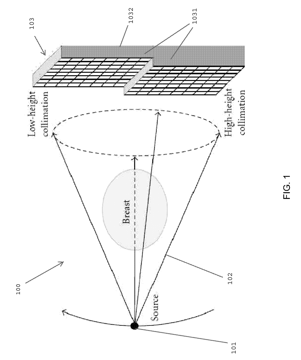
[0051]Included in embodiments of the invention are methods for extracting x-ray small-angle scattering data and using this segregated data to produce a high quality, high contrast x-ray image. A technique for extracting the x-ray small-angle scattering data involves collecting the x-ray projection data multiple times with varying collimation before an x-ray detector array. In preferred embodiments, each x-ray sum is acquired at least twice using different collimation aspect ratios. The projection data acquired with a collimator of a sufficiently large aspect ratio (otherwise referred to as a high collimation aspect ratio) contain mainly the primary beam with little scattering. In contrast, the co...


PUM


Abstract

The present invention relates to the field of x-ray imaging. More particularly, embodiments of the invention relate to methods, systems, and apparatus for imaging, which can be used in a wide range of applications, including medical imaging, security screening, and industrial non-destructive testing to name a few. Specifically provided as embodiments of the invention are systems for x-ray imaging comprising: a) a first collimator-and-detector assembly having a first operable configuration to provide at least one first dataset comprising primary x-ray signals as a majority component of its data capable of being presented as a first image of an object subjected to x-ray imaging; b) a second collimator-and-detector assembly having a second operable configuration or wherein the first collimator-and-detector assembly is adjustable to a second configuration to provide at least one second dataset comprising primary and dark-field x-ray signals as a majority component of its data capable of being presented as a second image of the object; and c) a computer operably coupled with the collimator-and-detector assemblies comprising a computer readable medium embedded with processing means for combining the first dataset and the second dataset to extract the dark-field x-ray signals and produce a target image having higher contrast quality than the images based on the first or second dataset alone. Such systems can be configured to comprise at least two collimator-and-detector assemblies or configurations differing with respect to collimator height, collimator aperture, imaging geometry, or distance between an object subjected to the imaging and the collimator-and-detector assembly.

Description

CROSS-REFERENCE TO RELATED APPLICATIONS[0001]This application relies on the disclosure of and claims the benefit of the filing date of U.S. Provisional Application No. 61 / 184,029, filed Jun. 4, 2009, the disclosure of which is incorporated by reference herein in its entirety.STATEMENT OF GOVERNMENT INTEREST[0002]This work was partially supported by the National Institutes of Health under Grants EB001685, EB006036, EB008476, CA135151 and CA127189. The U.S. Government has certain rights in the invention.BACKGROUND OF THE INVENTION[0003]1. Field of the Invention[0004]The present invention relates to the field of x-ray imaging. More particularly, embodiments of the invention relate to methods, systems, and apparatus for imaging, which can be used in a wide range of applications, including medical imaging, security screening, and industrial non-destructive testing to name a few.[0005]2. Description of the Related Art[0006]Since its invention in 1973, X-ray computed tomography (CT) has re...

Claims


Application Information

Patent Timeline
Patent Type & Authority Patents(United States)
IPC IPC(8): A61B6/00
CPCA61B6/032A61B6/4291A61B6/06A61B6/583
Inventor WANG, GECONG, WENXIANG
Owner VIRGINIA TECH INTPROP INC
Why Eureka
Try Eureka