
Preparation method of universal drug-loaded calcium phosphate cement (CPC) porous scaffold
A porous scaffold and bone cement technology, applied in the fields of medical science, tissue regeneration, prosthesis, etc., can solve the problem of difficult drug loading of calcium phosphorus bone cement
Patent Information
- Authority / Receiving Office
- CN · China
- Current Assignee / Owner
- Publication Date
- 2020-10-20
- Estimated Expiration
- Not applicable · inactive patent
Smart Images
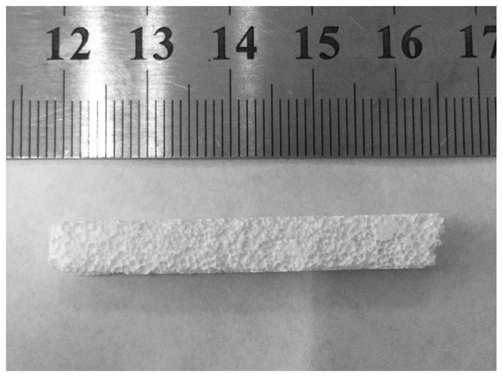
Figure 1 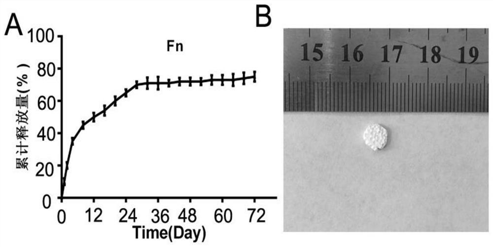
Figure 2 
Figure 3
Abstract
Description
technical field
[0001] The invention belongs to the field of biomedical materials, and relates to a preparation technology of a porous scaffold, in particular to a method for preparing a general-purpose drug-loaded calcium-phosphorus bone cement porous scaffold. Background technique
[0002] Bone is a dynamic tissue that constantly repairs and renews itself through remodeling. Bone tissue involves a variety of cells, signaling factors, growth factors, etc. Human bone tissue cells mainly include osteoblasts, osteoclasts, osteocytes, endosteal cells, vascular endothelial cells, and fibroblasts. The diameter of human osteoblasts is between 10-100um, which is differentiated from bone marrow mesenchymal stem cells. At present, bone tissue research mainly focuses on the research of biological scaffold materials, seed cells and factors.
[0003] Calcium phosphate bone cement is a non-ceramic hydroxyapatite, a bioactive bone repair material, usually composed of a liquid phase, a s...
Examples
preparation example Construction
[0030] A method for preparing a general-purpose drug-loaded calcium-phosphorus bone cement porous scaffold, the method steps are as follows:
[0031] (1) Prepare PLGA microspheres by emulsification method;
[0032] ⑵ Preparation of bone cement powder: tetracalcium phosphate Ca 4 (PO4) 2 O and anhydrous calcium hydrogen phosphate CaHPO 4 The powder is mixed to prepare CPC powder;
[0033] (3) Preparation of calcium-phosphorus bone cement microsphere mixed scaffold: After mixing CPC powder and solidification solution according to 10g: 3-10mL, mix evenly with PLGA microsphere and quickly fill it into a Φ5mm×5cm stainless steel abrasive tool, and grind one end flat After standing at room temperature for 2 minutes, remove the mold, quickly transfer to -80°C refrigerator to pre-freeze for 3-8 hours, and freeze-dry for more than 48 hours after complete freezing to obtain the mixed scaffold;
[0034] ⑷Preparation and sterilization of porous scaffolds: Soak the mixed scaffolds in a...
Embodiment 1
[0045] Tetracalcium phosphate Ca 4 (PO 4 ) 2 O(TTCP) and anhydrous calcium hydrogen phosphate CaHPO 4 (DCPA) powders were mixed at a molar ratio of 1:2 to prepare CPC powders. Mix CPC powder and sterile PBS solution according to the ratio of 10g: 3mL, mix evenly with 1gPLGA microspheres and fill it into a Φ5mm×5cm stainless steel abrasive tool, grind one end flat, pre-cure at room temperature for 2min, demould, and quickly transfer to Pre-freeze at -80°C for more than 5 hours, then freeze-dry for 48 hours after complete freezing. Then the hydrated bone cement was soaked in acetone at 4°C for 20 minutes, and then quickly washed with cold absolute ethanol, cold sterile water, and cold sterile PBS, dried at low temperature, and then sterilized with an ultraviolet lamp for use. It is measured that the pores of the support are interconnected with each other, the pore size is 150-400 μm, and the porosity is 43.5% ( figure 1 ).
Embodiment 2
[0047] Tetracalcium phosphate Ca 4 (PO 4 ) 2 O(TTCP) and anhydrous calcium hydrogen phosphate CaHPO 4 (DCPA) powders were mixed at a molar ratio of 1:2 to prepare CPC powders. Mix CPC powder and Fn PBS solution according to 10g: 3mL, mix evenly with 1g PLGA microspheres and fill it into a Φ5mm×5cm stainless steel abrasive tool, grind one end flat, pre-cure at room temperature for 2min, demould, and quickly transfer to - Pre-freeze in the refrigerator at 80°C for more than 5 hours, and freeze-dry for 48 hours after complete freezing. Then the hydrated bone cement was soaked in acetone at 4°C for 20 minutes, and then quickly washed with cold absolute ethanol, cold sterile water, and cold sterile PBS, dried at low temperature, and then sterilized with an ultraviolet lamp for use. The Fn detection kit was used to detect the release of fibronectin and the release curve of Fn was drawn ( figure 2 ).