
Injectable composit material for bone repair, and preparation method thereof
a composite material and bone technology, applied in the field of material science, can solve the problems of increasing the recovery time of patients, increasing the duration and cost of surgery, and the disadvantages of autologous bone, and achieve the effects of reducing the risk of infection
Patent Information
- Authority / Receiving Office
- US · United States
- Patent Type
- Applications(United States)
- Current Assignee / Owner
- Publication Date
- 2018-09-20
Smart Images
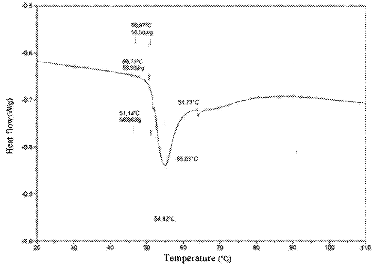
Figure 1 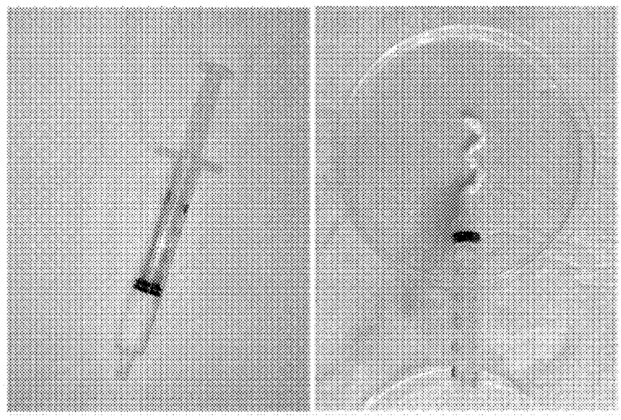
Figure 2 
Figure 3
Abstract
Description
TECHNICAL FIELD
[0001] The present invention belongs to the field of material science, and particularly to an injectable composite material for bone repair and the preparation method thereof. The composite material can be widely used in the field of orthopaedics and plastic surgery.BACKGROUND
[0002] The repair and reconstruction of impaired bone tissues are important tasks that orthopaedic surgeons need to face all over the world. Bone grafting materials are needed in the repair of these bone tissues. Autologous bone tissues, i.e., healthy bone tissues harvested from the patient's own body, have been considered worldwide the golden standard for the repair of impaired bone tissues. The autologous bone has all the properties required for repairing bone tissues, such as excellent osteoinductivity, osteoconductivity, osteogenecity, and safety. However, there are clinically unavoidable disadvantages when using autologous bones. Since bone tissues need to be harvested from the donor site of t...
Examples
example 1
The Preparation and Characterization of Biological Tissue Matrix / Calcium Hydrophosphate Composite Material
[0059]1) A microfibrillar biological tissue matrix material was weighted, which was obtained from decellularized and antigen-extracted porcine dermis and sterilized by Co60 gamma rays. The material was suspended in 0.9% of physiological saline in a concentration of 10 mg / mL.
[0060]2) The material was washed 3 or more times with sterile deionized water. The parameters of the prepared decellularized matrix microfiber were shown in FIG. 1. It has an average aspect ratio of 0.05-0.95, and a particle size distribution of 40-1000 μm.
[0061]3) Calcium hydrophosphate microparticles were weighted and sterile deionized water was added to prepare a 10 mg / mL of homogeneous suspension.
[0062]4) The calcium hydrophosphate microparticles were rapidly passed through a filter membrane with a pore diameter of 40 micron and the oversized agglomerated particles therein were removed.
[0063]5) The suspen...
example 2
Bone Defect Repair Experiment of the Injectable Biological Tissue Matrix / Calcium Hydrophosphate Composite Material
[0071]The injectable biological tissue matrix / calcium hydrophosphate composite material prepared according to the present application was used to evaluate the bone forming ability of this material with a mouse calvarial defect model of nude mouse without thymus. The mice (6 cases) were anesthetized and then the head furs thereof were shaved off. A 1 centimeter skin incision was cut along the sagittal suture of the mouse head. Circular defect with a diameter of 3.5 millimeter was made on the skull with a dental drill after putting the skin apart. The bone defect was filled with the material prepared according to the present invention, and then the wound was sutured. Six weeks later, the mice were sacrificed humanely. The skull after repairing was observed grossly. It is found that the material and the surrounding bone tissue integrated well. X-ray observation shows that a...
example 3
Bone Defect Repair Experiment of the Injectable Biological Tissue Matrix / Calcium Hydrophosphate Composite Material as a Carrier for Fresh Bone Marrow
[0072]The injectable biological tissue matrix / calcium hydrophosphate composite material prepared in Example 1 was mixed with fresh mouse bone marrow aspirate to prepare a paste-like material. The mouse model used in Example 2 was used. The bone defects were filled with the composite material loaded with fresh bone marrow aspirate, and then the wounds were sutured. Six weeks later, X-ray observations revealed that the entire bone defect was filled with newly formed bone tissue. No tissue gap was found at the edge of the bone defects. Micro-CT scanning showed the same results that the entire bone defect had been filled with newly formed bone tissues, and a seamless connection was formed between the new bone and the implant. Quantitative analysis of bone formation showed that the material prepared in Example 1 could form much more bones th...