
Fabp4 as a therapeutic target in skin diseases
Patent Information
- Authority / Receiving Office
- US · United States
- Current Assignee / Owner
- Publication Date
- 2020-05-07
Smart Images
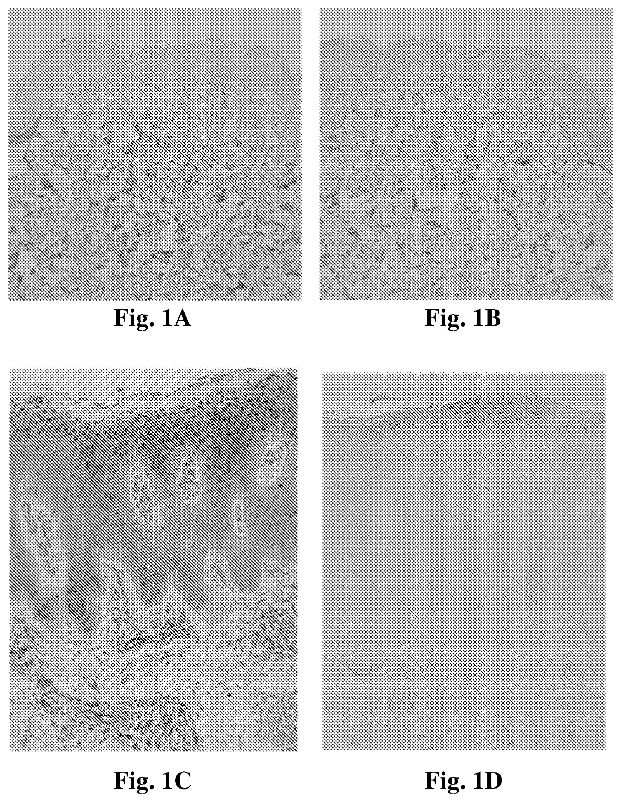
Figure 1 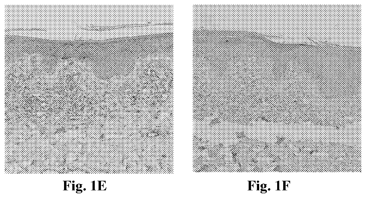
Figure 2 
Figure 3
Abstract
Description
SEQUENCE LISTING
[0001] The Sequence Listing submitted in text format (.txt) filed on Jan. 7, 2020, named “SequenceListing.txt”, created on Dec. 16, 2019 (2.36 KB), is incorporated herein by reference.TECHNOLOGICAL FIELD
[0002] The present invention relates to methods of regulating proliferation and / or differentiation of keratinocytes and immune cells, more specifically to methods of treating pathologies characterized by hyperproliferative keratinocytes or inflammatory skin diseases.BACKGROUND ART
[0003] References considered to be relevant as background to the presently disclosed subject matter are listed below:
[0004] [1] Furuhashi et al., Nat Rev Drug Discov 2008, 7(6), 489-503
[0005] [2] Coe et al., Biochim Biophys Acta 1998, 1391(3), 287-306
[0006] [3] Hotamisligil et al., Science 1996, 274(5291), 1377-1379
[0007] [4] Maeda et al., Cell Metab 2005, 1(2), 107-119
[0008] [5] Furuhashi et al., Nature 2007, 447(7147), 959-965
[0009] [6] Tuncman et al., PNAS 2006, 103(18), 6970-6975
[0010] [7] Garin-Sh...
Examples
example 1
Tissue Expression Analysis of FABP4, FABPS, and PPARγ in Human Psoriatic Skin Lesions
[0117]Punch biopsies (4 mm in diameter) were obtained from disease-involved skin of patients with psoriasis (n=10). Biopsies were further obtained from involved skin of patients with chronic dermatitis (n=5). Normal skin was obtained from patients after surgical reduction of redundant skin (n=10). The tissues were fixed in formalin and embedded in paraffin. For histopathological examination with light microscopy, the sections were stained with hematoxylin and eosin (H&E) and observed by a pathologist who confirmed the diagnosis. In addition, for immunohistochemical analysis, the sections were stained with the following antibodies: FABP4 (Rabbit polyclonal anti FABP4 antibody, PAB12276, Abnova), FABPS (Rabbit polyclonal anti FABPS antibody, SC-50379, Santa Crus) and PPARγ (mouse monoclonal anti-PPARγ antibody, E-8, Santa Crus), all diluted 1:50. The paraffin-embedded and cryopreserved tissues were pr...
example 2
Tissue Expression Analysis of FABP4 in Human Cutaneous T Cell Lymphoma (CTCL) Skin Lesions
[0123]CTCL is a group of neoplasms of skin-homing T cells. Mycosis fungoides (MF) represents the most common type of CTCL and accounts for approximately 50% of all primary cutaneous lymphomas. Although malignant in nature, MF has a protracted clinical course with inflammatory, dermatitis-like presentation [8].
[0124]The expression of FABP4 among patients with MF was studied by performing an immunohistochemical analysis of lesional skin. Samples from 5 patients with MF were tested and compared to normal human skin. Punch biopsies (6 mm in diameter) were obtained from patients with MF, from disease-involved skin. Normal skin was obtained from patients after surgical reduction of redundant skin. Tissue sections were processed and stained with anti-FABP4 antibody as described in Example 1.
[0125]High expression levels of FABP4 were observed in MF lesions compared to normal skin with all tested patien...
example 3
Overexpressing FABP4 in Primary Murine Keratinocytes
[0128]The introduction of FABP4 into keratinocytes was suggested to create a hyperproliferative state with impaired differentiation as seen in psoriasis. Differentiation was assessed by measuring the expression of two keratinocyte differentiation markers K1 and K5. K5 is a keratin whose level does not change during keratinocyte differentiation and therefore serves as a loading control, while K1 is induced during normal keratinocyte differentiation.
[0129]For assessing the effect of FABP4 overexpression in mouse primary keratinocytes, cells were infected with a lentiviral vector construct containing the FABP4 gene. The FABP4 lentiviral vector, named FABP4-T2A-EGFP, was constructed with the FABP4 gene under the CMV promotor, and contained the green fluorescence protein (GFP) gene as a reporter of expression. The presence of a T2A residue at the N-terminus of the FABP4 protein precludes FABP4 detection by anti-FABP4 commercial antibodi...