X-ray computerized tomography system and method

An X-ray and computer technology, applied in the field of X-ray computed tomography system, can solve the problems of not changing the opening of the collimator, not adjusting the exposure area of ​​the object to be inspected, and not considering the size change and shape change of the ROI, so as to reduce the X-ray Effects of radiation dose, reduction of additional exposure, and correction of opening width

Active Publication Date: 2014-01-22
SIEMENS SHANGHAI MEDICAL EQUIP LTD
View PDF6 Cites 0 Cited by
  • Summary
  • Abstract
  • Description
  • Claims
  • Application Information

AI Technical Summary

Problems solved by technology

However, this method only proposes a solution for using a fixed FOM in a single rotation scan, without considering the size and shape changes of the ROI in the scanning direction, where the scanning direction is the horizontal direction of the examination bed in and out of the rack in the CT system, Perpendicular to the phi plane, usually in the z-axis direction
[0005] The patent application whose publication number is CN 1446517A discloses an X-ray data acquisition system, which adopts an elliptical cross section or a circular cross section, and calculates the transmission value according to the target value of the standard deviation of the image, the expected value of the standard deviation of the image and the thickness factor. The dose value of X-rays within the predetermined angle range of the body axis direction of the object to be inspected is achieved by changing the tube current of the X-ray tube, but the opening of the collimator is not changed, nor is it scanned according to the object to be inspected The size of the direction changes to adjust the exposure area of ​​the object to be inspected accordingly

Method used


Image

  • X-ray computerized tomography system and method
  • X-ray computerized tomography system and method
  • X-ray computerized tomography system and method

Examples


Embodiment Construction

[0018] In order to make the purpose, technical solution and advantages of the present invention clearer, the following examples are given to further describe the present invention in detail.

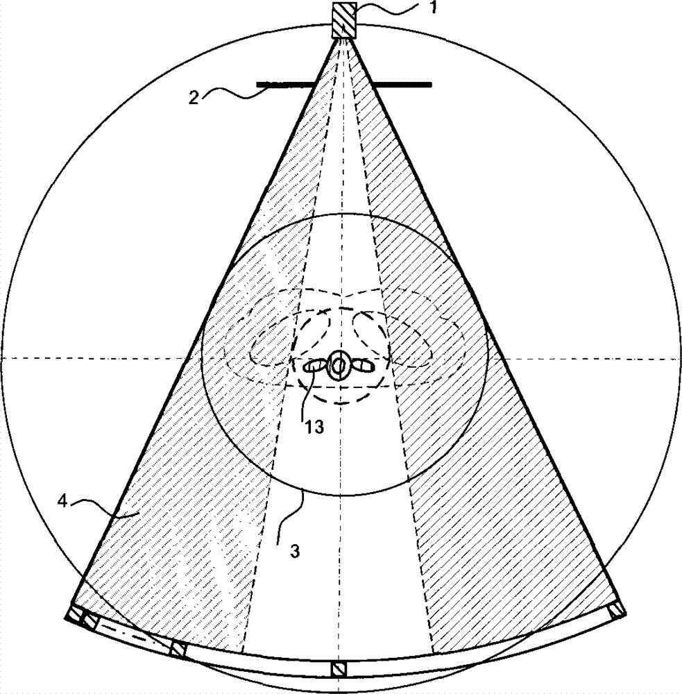
[0019] The present invention calculates the opening width of the collimator according to the different sizes of multiple circular sections of the object to be inspected in the scanning direction, and adjusts the opening of the collimator according to the opening width, so as to perform CT scanning on the object to be inspected , in order to reduce the additional exposure of the area around the object to be examined, while reducing the X-ray dose received by the patient.

[0020] In the present invention, the object to be inspected may be a certain area of ​​the human body, or a certain organ or tissue of the human body. In the embodiment of the present invention, the object to be inspected is the spine, and the scanning direction (that is, the horizontal direction in which the examinatio...


PUM


Abstract

The invention discloses an X-ray computerized tomography system and an X-ray computerized tomography method. The system comprises an X-ray tube, a collimator, a detector, a computing assembly and an adjusting assembly, and is used for shooting a plurality of circular sections of an object to be detected in the scanning direction, wherein the detector comprises a plurality of detector channels; the X-ray tube, the collimator and the detector rotate synchronously around the object to be detected; the collimator is a collimator with an adjustable opening; the computing assembly is used for computing the opening width of the collimator needed for irradiating each circular section respectively, and transmitting the opening width to the adjusting assembly; and the adjusting assembly is used for adjusting the opening of the collimator according to the opening width. The invention further discloses the X-ray computerized tomography method. Due to the adoption of the system and the method, extra exposure of the surrounding area of the objected to be detected can be reduced, and the X-ray dosage received by a patient can be reduced.

Description

technical field [0001] The invention relates to the field of medical imaging, in particular to an X-ray computed tomography system and method. Background technique [0002] In X-ray computed tomography (CT) system, dose characteristic is a very important performance index. Keeping the scanning dose as low as possible has become a general rule in the development of current CT systems. In a CT system, the field of measurement (FOM) is defined as the cross-sectional area where the object to be inspected gets full radiation when the X-ray tube rotates 360 degrees in a single rotation scan, which determines the direct detection of the object to be inspected. The scan dose applied and the size of the X-ray irradiated area. [0003] At present, most CT systems use a fixed circular FOM and a phi collimator with a constant opening (that is, a fan beam collimator) to scan the object to be inspected, where the phi plane is the plane where the cross-section of the human body is locate...

Claims


Application Information

Patent Timeline
Patent Type & Authority Patents(China)
IPC IPC(8): A61B6/03
Inventor 田毅陈马昊
Owner SIEMENS SHANGHAI MEDICAL EQUIP LTD
Why Eureka
Try Eureka