Ultrasonic diagnostic device, and method for measuring intima-media complex thickness

A diagnostic device, ultrasonic technology, applied in the direction of acoustic wave diagnosis, infrasonic wave diagnosis, ultrasonic/sonic wave/infrasonic wave diagnosis, etc.

Inactive Publication Date: 2012-05-23
KONICA MINOLTA INC
View PDF8 Cites 11 Cited by
  • Summary
  • Abstract
  • Description
  • Claims
  • Application Information

AI Technical Summary

Problems solved by technology

[0011] In the ultrasonic diagnostic apparatus of the conventional example, when it is desired to measure the state of the carotid artery as the detection target, only skilled persons who have accumulated sufficient training can perform accurate measurement.
[0013] Even if an image showing a state in which the carotid artery can be accurately measured can be displayed by touching the probe at an appropriate position, it is difficult to perform measurement while maintaining the correct position because the operator manipulates the probe by hand.

Method used


Image

  • Ultrasonic diagnostic device, and method for measuring intima-media complex thickness
  • Ultrasonic diagnostic device, and method for measuring intima-media complex thickness
  • Ultrasonic diagnostic device, and method for measuring intima-media complex thickness

Examples


Embodiment approach 1

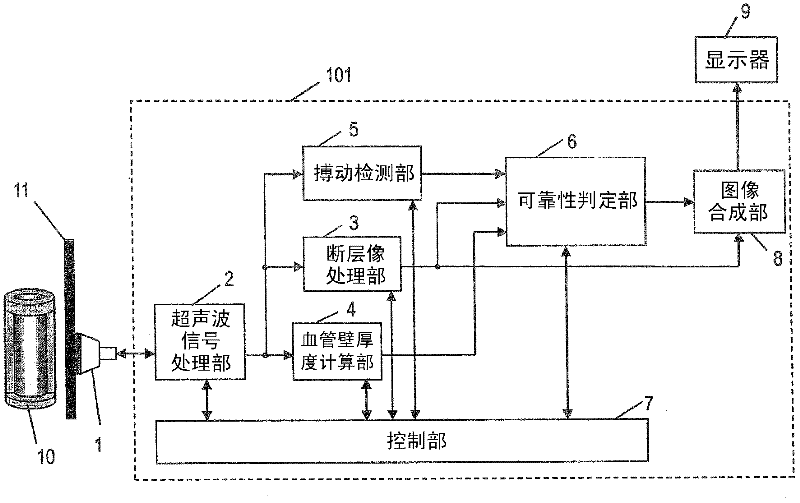
[0064] figure 1 It is a block diagram of the ultrasonic diagnostic apparatus according to Embodiment 1 of the present invention. The ultrasonic diagnostic apparatus 101 of this embodiment includes: an ultrasonic signal processing unit 2 ; a tomographic image processing unit 3 ; a blood vessel wall thickness calculation unit 4 ; a pulsation detection unit 5 ; a reliability determination unit 6 ;

[0065]The probe 1 has an ultrasonic vibrator, transmits ultrasonic waves to the subject through the ultrasonic vibrator, receives reflected ultrasonic waves from the subject, and converts them into electrical signals. The ultrasonic signal processing unit 2 is configured so that the probe 1 can be detached, supplies a drive pulse to the ultrasonic vibrator of the probe 1 at a predetermined timing, drives the probe 1 to transmit ultrasonic waves, and performs transmission processing. In addition, an electric signal is received from the probe 1, reception processing necessary for cons...

Embodiment approach 2

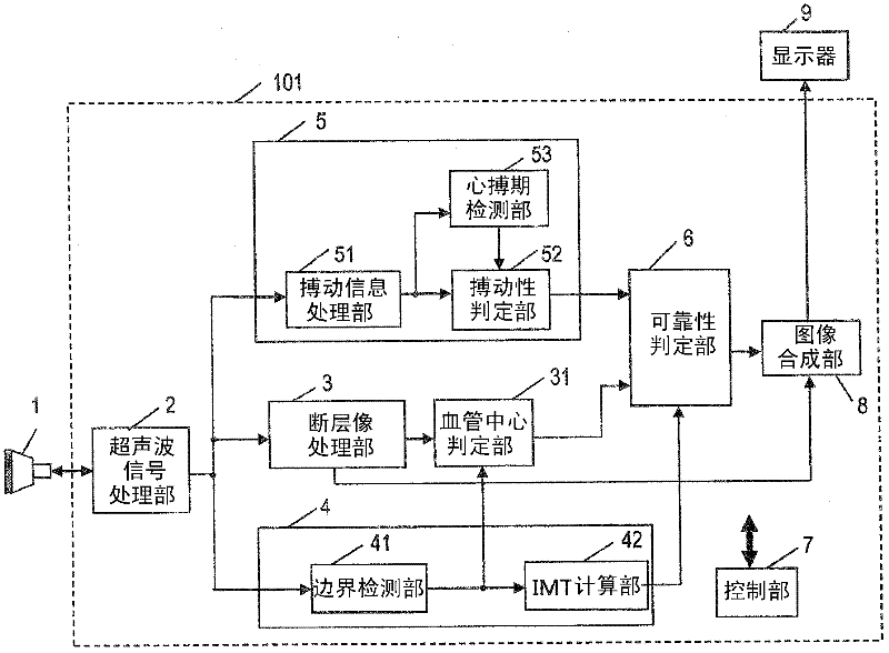
[0134] Next, for Embodiment 2, use Figure 11 to explain. Also, a typical action flow chart is the same as Figure 5 same.

[0135] Figure 11 It is a block diagram of an ultrasonic diagnostic apparatus according to Embodiment 2 of the present invention. The ultrasonic diagnostic apparatus 102 of this embodiment includes: an ultrasonic signal processing unit 2 ; a tomographic image processing unit 3 ; a blood vessel wall thickness calculation unit 4 ; a pulsation detection unit 50 ; a reliability determination unit 6 ;

[0136] with use figure 2 The difference of Embodiment 1 described above is that a pulsation detection unit 50 is provided instead of the pulsation detection unit 5, and the pulsation detection unit 50 has: a pulsation information processing unit 51; a pulsation determination unit 52; an ECG signal processing unit 54 and a Periodically detect hand 55.

[0137] Since the pulsation information processing unit 51 and the pulsation determination unit 52 have...

Embodiment approach 3

[0147] Next, use Figure 12 and Figure 13 , Embodiment 3 of the present invention will be described. Figure 12 is a block diagram of an ultrasonic diagnostic apparatus according to Embodiment 3 of the present invention, Figure 13 It is a flowchart showing a typical operation of Embodiment 3 of the present invention.

[0148] The ultrasonic diagnostic apparatus 103 according to Embodiment 3 includes: an ultrasonic signal processing unit 2; a tomographic image processing unit 3; a blood vessel wall thickness calculation unit 4; a pulsation detection unit 5; a reliability determination unit 6; a control unit 7; The long axis determination unit 20 ; the stability determination unit 21 and the blood vessel center determination unit 31 .

[0149] The difference from Embodiment 1 is that a major axis determination unit 20 and a stability determination unit 21 are provided, and the reliability determination unit 6 uses the determination results of the major axis determination un...


PUM


Abstract

Disclosed is an ultrasonic diagnostic device provided with: an ultrasonic wave signal processing unit which performs a transmission processing for transmitting ultrasonic waves to the blood vessel of a subject by driving a probe, and performs a reception processing for generating a reception signal from the ultrasonic wave which reflected from the blood vessel of the subject and which was received by the probe; a tomographic image processing unit which creates a tomographic image from the reception signal; an interface detection unit which detects the lumen-intima interface and media-adventitia interface of the blood vessel from the reception signal or the tomographic image; a blood vessel wall thickness calculating unit which calculates the distance between the lumen-intima interface and the media-adventitia interface, which were detected by means of the interface detection unit, as the blood vessel wall thickness value; a reliability determination unit which determines the reliability of the blood vessel wall thickness value on the basis of the features on an image information or on a signal at the lumen-intima interface and media-adventitia interface positions, which were detected by means of the reception signal or the tomographic image; and a control unit which determines, on the basis of the determination results from the reliability determination unit, that the blood vessel wall thickness value is the intima-media complex thickness.

Description

technical field [0001] The present invention relates to an ultrasonic diagnostic device and a method for measuring the thickness of an intima-media complex using the same. Background technique [0002] In the diagnosis of arteriosclerosis using an ultrasonic diagnostic apparatus, it is known that the intima-media thickness (IMT: Intima-Media Thickness, hereinafter abbreviated as IMT) of the carotid artery is an important index for identifying the initial stage of atherosclerosis. IMT refers to the thickness of the complex of intima and media in the vessel wall of the carotid artery. Intima-media complexes, such as Figure 18 As shown, the layer seen between the lumen and the adventitia of the blood vessel is detected by inspection, the boundary between the lumen and the intima of the blood vessel (hereinafter referred to as the lumen-intima boundary) and the boundary between the media and the adventitia (hereinafter referred to as the media-adventitia boundary), and its thi...

Claims


Application Information

Patent Timeline
Patent Type & Authority Applications(China)
IPC IPC(8): A61B8/06
CPCA61B8/5284A61B8/0858A61B8/0891
Inventor 福元刚智川端章裕占部真树子铃木隆夫西村有史
Owner KONICA MINOLTA INC
Why Eureka
Try Eureka