Automatic vessel wall edge detection method based on intravascular ultrasound image sequence

An ultrasonic image, automatic detection technology, applied in image analysis, image data processing, instruments, etc., can solve problems such as poor extraction effect, large dependence on initial edge position, and difficulty in convergence.

Inactive Publication Date: 2013-12-18
BEIJING UNIV OF TECH
View PDF1 Cites 11 Cited by
  • Summary
  • Abstract
  • Description
  • Claims
  • Application Information

AI Technical Summary

Problems solved by technology

These algorithms need to manually select the initial contour, have poor anti-noise ability, and have the disadvantages that the boundary depression is not easy to converge
In order to improve the curve convergence characteristics of the closed boundary depression, the GVF-Snake model, which is de...

Method used


Image

  • Automatic vessel wall edge detection method based on intravascular ultrasound image sequence
  • Automatic vessel wall edge detection method based on intravascular ultrasound image sequence
  • Automatic vessel wall edge detection method based on intravascular ultrasound image sequence

Examples


Embodiment Construction

[0056] An automatic edge detection method based on intravascular ultrasound, the specific steps are as follows:

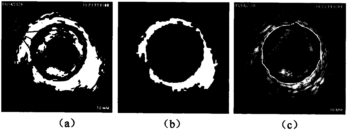
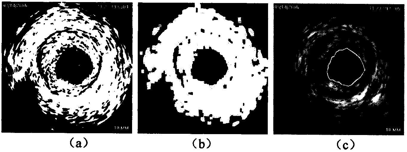
[0057] Step 1. According to the characteristics of the intravascular ultrasound image, the image processing method is comprehensively used to pre-extract the outer edge of the blood vessel wall to obtain the initial edge of the blood vessel wall.

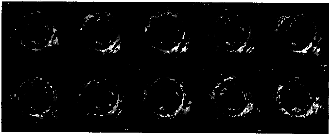
[0058] Step 1.1, such as figure 1 As shown in , select 10 consecutive frames of images, calculate their time variance map, and use the continuity of the frames to remove some noise (such as halo artifacts, etc.) and some breakpoints at the connection edge ( Such as the sound shadow area):

[0059] V ( x , y ) = 1 n - 1 Σ m = 1 n [ ...


PUM


Abstract

An automatic vessel wall edge detection method based on an intravascular ultrasound image sequence includes the steps of carrying out coarse detection, obtaining an initial edge of a first frame image by means of series of image processing methods according to information characteristics on a vessel wall, carrying out fine detection, improving the GVF-snake algorithm, leading in self-adjusting factors and self-adaptation external force, improving the gradient influences of data items close to the edge, reducing the smooth effect caused by diffusion, meanwhile adjusting the direction of force according to the relative position of the image edge and a current outline curve, enlarging the acquisition range of a moving outline, and solving the problems that a local area can not be reached, and accordingly the required edge can not be obtained so that the initial edge can be precisely converged to a practical vessel wall edge. In the deforming process, spline B is added for three times so as to reduce control points, improve convergence speed, achieve fairing of the outline curve and obtain the final edge of the first frame image. Due to the fact that time and space correlation between adjacent images is large, the obtained final edge serves as the initial edge of an image of the next frame, fine detection is repeated, and similarly, the vessel wall edge of the image sequence is obtained.

Description

technical field [0001] The invention relates to an automatic detection method of blood vessel edge based on internal ultrasonic image sequence, which belongs to the field of computer medical image analysis. , The accurate measurement of blood vessel thickness is conducive to the discovery or identification of early coronary atherosclerotic plaques, which has important clinical significance. Background technique [0002] Worldwide, cardiovascular and cerebrovascular diseases have become the leading cause of human death. Intravascular Ultrasound (IVUS) is the latest technology for the diagnosis of cardiovascular and cerebrovascular diseases. Extracting the inner and outer edges of the blood vessel wall in intravascular ultrasound images, and image analysis based on this, has important clinical significance for discovering or identifying early coronary atherosclerotic plaques. [0003] In recent years, vessel edge detection algorithms based on intravascular ultrasound images ...

Claims


Application Information

Patent Timeline
IPC IPC(8): G06T7/00
Inventor 汪友生张丽杰吴焕焕王志东
Owner BEIJING UNIV OF TECH
Why Eureka
Try Eureka