Arteriovenous retinal blood vessel classification method based on eye fundus image

A technology of retinal blood vessels and fundus images, which is applied in the field of arteriovenous retinal blood vessel classification based on fundus images, can solve the problems of low degree of automation and achieve improved classification results without manual intervention and high classification accuracy

Active Publication Date: 2015-04-29
杭州求是创新健康科技有限公司
View PDF3 Cites 30 Cited by
  • Summary
  • Abstract
  • Description
  • Claims
  • Application Information

AI Technical Summary

Problems solved by technology

[0007] Blood vessel segmentation, optic disc positioning and vessel classification (arteriovenous split) on fundus images are the basis of retinal vess

Method used


Image

  • Arteriovenous retinal blood vessel classification method based on eye fundus image
  • Arteriovenous retinal blood vessel classification method based on eye fundus image
  • Arteriovenous retinal blood vessel classification method based on eye fundus image

Examples


Embodiment Construction

[0066] The present invention will be described in detail below with reference to the drawings and specific embodiments.

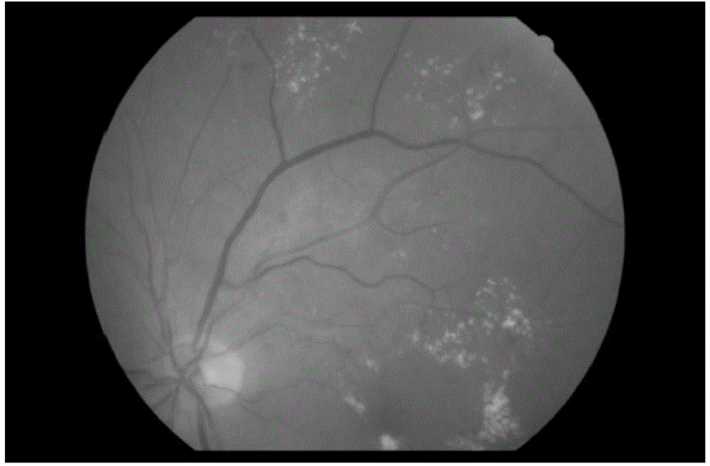
[0067] This embodiment takes figure 1 The fundus image shown is taken as an example to illustrate the arteriovenous retina vessel classification method based on the fundus image, and the size of the fundus image is 3000×3000. There are bright rings in the fundus image due to the ring reflection caused by photography, the non-vascular step edge around the optic disc, patchy lesions, and hemorrhagic lesions.

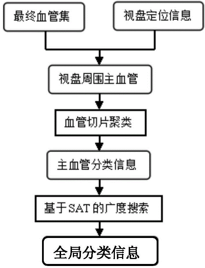
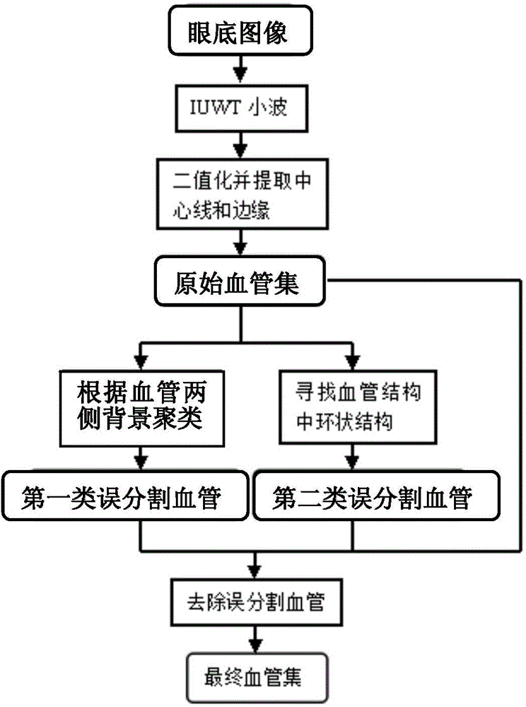
[0068] The arteriovenous retinal vessel classification is performed on the fundus image, and the classification process is as follows: figure 2 shown, including the following steps:

[0069] (1) Obtain the global blood vessel set (i.e. the final blood vessel set) and the optic disc positioning information of the fundus image, the global blood vessel set is the collection of all blood vessels in the fundus image, and the optic disc positioning informa...


PUM


Abstract

The invention discloses an arteriovenous retinal blood vessel classification method based on an eye fundus image. The method includes the steps that first, a global blood vessel set and optic disk positioning information of the fundus image are acquired, the global blood vessel set is a set of all blood vessels in the fundus image, and the optic disk positioning information comprises the optic disk center of the fundus image; second, main blood vessels are determined according to the global blood vessel set and the optic disk positioning information and classified so that main blood vessel classification information can be obtained; third, the main blood vessel classification information is used for classifying the blood vessels in the global blood vessel set through a breadth first-search algorithm based on SAT so that global classification information can be obtained. According to the method, the classification information of the main blood vessels around an optic disk is first obtained, external expansion diffusion is performed from the main blood vessels through the breadth first-search algorithm based on SAT so that all the blood vessels can be obtained, a complete automatic blood vessel classification method is achieved, manual intervention is not needed, and classification precision is high.

Description

technical field [0001] The invention relates to the technical field of computer-aided diagnosis, in particular to a method for classifying arteriovenous retinal blood vessels based on fundus images. Background technique [0002] With the rapid development of the field of artificial intelligence in computer technology, computer-aided diagnosis technology is also gradually developed. Computer-aided diagnosis technology refers to the use of imaging, medical image processing technology and other possible physiological and biochemical means, combined with computer analysis and calculation, to assist radiologists in finding lesions and improving the accuracy of diagnosis. [0003] Usually, computer-aided diagnosis in medical imaging is divided into three steps, as follows: the first step is to extract the lesion from the normal structure; the second step is to quantify the image features; the third step is to process the data and draw conclusions . [0004] Because the computer ...

Claims


Application Information

Patent Timeline
IPC IPC(8): G06K9/62A61B3/12
CPCA61B3/12G06V10/457G06F18/23213G06F18/24
Inventor 吴健黎罗河邓水光李莹尹建伟吴朝晖
Owner 杭州求是创新健康科技有限公司
Why Eureka
Try Eureka