Angiography image acquisition device and method

An image acquisition and angiography technology, applied in the field of X-ray display, can solve problems such as affecting the operation process of the surgeon

Active Publication Date: 2015-06-24
乐普(北京)医疗装备有限公司
View PDF6 Cites 1 Cited by
  • Summary
  • Abstract
  • Description
  • Claims
  • Application Information

AI Technical Summary

Problems solved by technology

After wearing stereoscopic glasses, it will affect the operation process of the surgeon. Therefore, it is necessa

Method used


Image

  • Angiography image acquisition device and method
  • Angiography image acquisition device and method
  • Angiography image acquisition device and method

Examples


Embodiment Construction

[0051] The present invention will be described in further detail below in conjunction with the accompanying drawings and embodiments. The following examples are used to illustrate the present invention, but should not be used to limit the scope of the present invention.

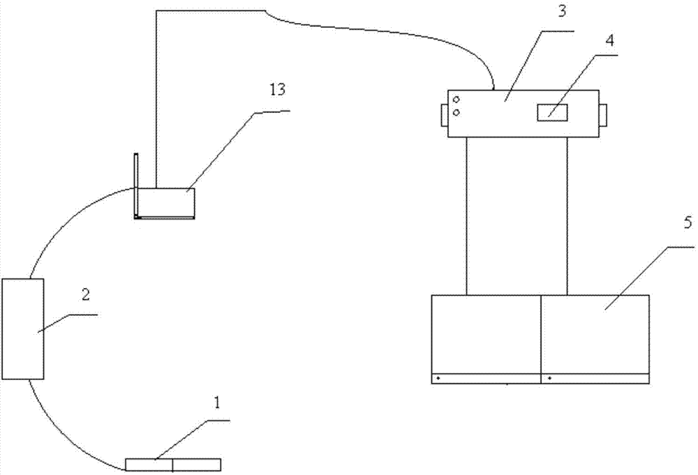
[0052] The invention provides an angiographic image acquisition device, such as figure 1 , Figure 4 As shown, the device includes:

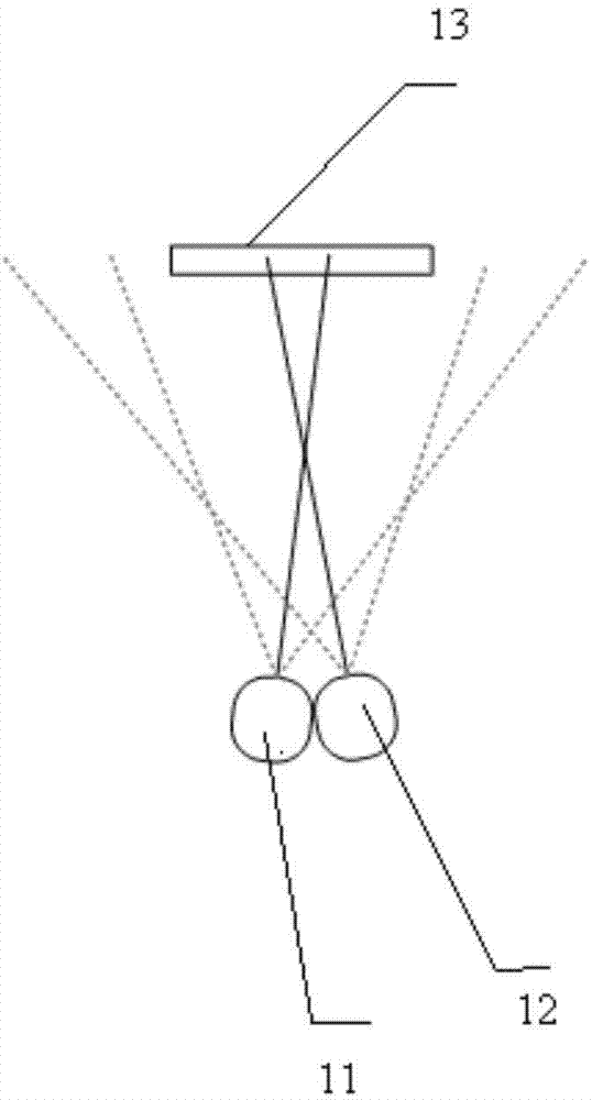
[0053] The X-ray imaging unit 1 comprises a left X-ray tube 11, a right X-ray tube 12, and a flat panel detector 13, the left X-ray tube 11 and the right X-ray tube 12 are placed side by side, and the flat panel detector 13 Placed parallel to the two X-ray tubes at a certain distance, the left X-ray tube 11 and the right X-ray tube 12 transmit the collected image signals to the flat panel detector 13 in time division, and the flat panel detector 13 Generate a first original image and a second original image respectively, and send the first original image and the second origi...


PUM


Abstract

The invention discloses an angiography image acquisition device and method. The device comprises an X-ray imaging unit, an image processing unit, a display unit, an image storage unit and a control unit, wherein the X-ray imaging unit is used for generating raw images of X-ray and sending the raw images to the image processing unit which calculates 3D fusion parameters; the display unit receives the first raw image, the second raw image and the 3D fusion parameters and conducts display through a 3D stereoscopic image form; the image storage unit conducts storage on the raw images and the control unit provides control pulses for the X-ray imaging unit and controls the angle of an X-ray bulb tube simultaneous. By means of the angiography image acquisition device and method, 3D stereoscopic images are generated in real time, the display effect of CT/RM 3D reconstruction is achieved simultaneously, the image overlapping influence is reduced, specific structure space location relationship is provided, and image lines can be displayed more clearly and visually.

Description

technical field [0001] The present invention relates to the technical field of X-ray display, and more particularly relates to an angiographic image acquisition device and method. Background technique [0002] Cardiovascular disease remains the leading cause of death in most developed countries. For patients presenting with symptoms of cardiovascular disease, the primary diagnostic and therapeutic approach is usually via interventional medicine. Catheters are inserted through arteries or veins into the ventricle. A catheter is used to inject a contrast agent into blood vessels so that the blood vessels can be visualized under X-rays. This procedure is called angiography. [0003] CT / MR three-dimensional reconstruction technology (3D-Reconstruction) refers to helical scanning and three-dimensional reconstruction of the observation site to generate three-dimensional reconstruction images that can be observed from multiple directions. CT / MR three-dimensional reconstruction t...

Claims


Application Information

Patent Timeline
IPC IPC(8): A61B6/03
Inventor 孙晓伟邝凯毅
Owner 乐普(北京)医疗装备有限公司
Why Eureka
Try Eureka