Vaginal and cervical examination instrument

A technology of cervical inspection and treatment instrument, applied in colposcopy, medical science, endoscopy, etc., can solve the problems of large economic burden, time-consuming and labor-intensive, lost treatment opportunities, etc.

Active Publication Date: 2018-05-22
GUANGZHOU PULU MEDICAL TECH CO LTD +2
View PDF10 Cites 0 Cited by
  • Summary
  • Abstract
  • Description
  • Claims
  • Application Information

AI Technical Summary

Problems solved by technology

[0002] At present, gynecological examination needs to go to the hospital to be checked by professional and technical personnel, which is time-consuming and laborious, and at the same time requires a great economic burden
In gynecological diseases, cervical cancer, cervicitis, cervical polyps, cervical cysts, and vaginitis, etc., if you go to check after the symptoms appear, the disease has developed to a certain extent at this time, and it is very likely that you will lose the best treatment. Chance

Method used


Image

  • Vaginal and cervical examination instrument
  • Vaginal and cervical examination instrument
  • Vaginal and cervical examination instrument

Examples


Embodiment Construction

[0030] Embodiments of the present invention are described in detail below:

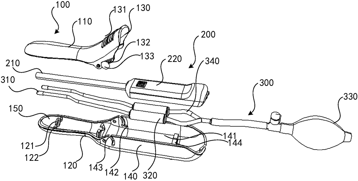
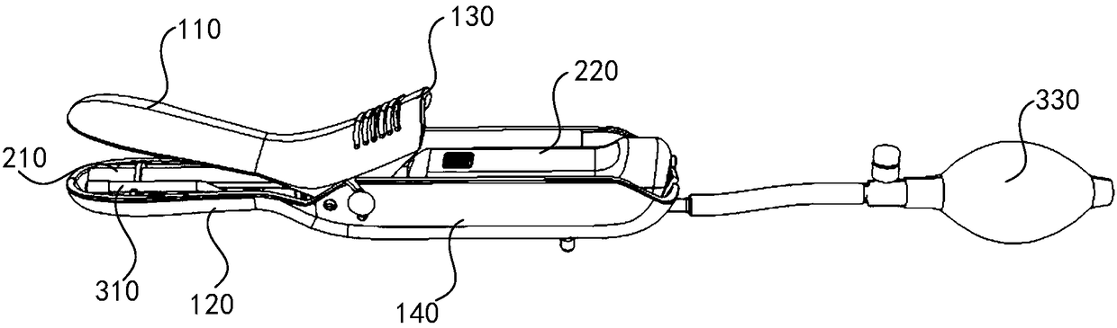
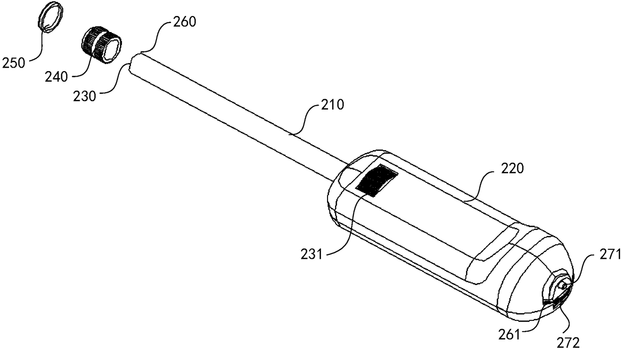
[0031] Such as Figure 1-2 As shown, a vagina and cervix examination instrument includes a vagina dilator 100 and an endoscope 200 . The vaginal dilator 100 includes a first expansion piece 110 and a second expansion piece 120 arranged up and down oppositely. The first expansion piece 110 and the second expansion piece 120 are hinged to each other, and the first expansion piece 110 and the second expansion piece 120 cooperate to form an operation channel 150 . The endoscope 200 is provided with a wireless transmission module (not shown in the drawings) and a camera located in the operation channel 150 . The wireless transmission module is a WiFi transmission module, a Bluetooth transmission module, and the like. The camera head is located at the end of the endoscope 200 and close to the end of the second dilating piece 120 protruding into the vagina. By extending the dilator 100 into the vagina, t...


PUM


Abstract

The invention discloses an instrument for examining and treating vagina and cervix, which includes a vagina dilator and an endoscope. The second expansion pieces are hinged to each other and the cooperation between the first expansion piece and the second expansion piece forms an operation channel, and the endoscope is provided with a wireless transmission module and a camera located in the operation channel. The invention can connect the photographed image with the external display terminal (mobile phone, tablet computer, etc.) through the wireless transmission module, and upload and store the inspection result in time. The operation is simple, time-saving and labor-saving, and the structure is simple, and the occupied space is small, so that the patient can perform vaginal examination at home. In addition, the camera of the present invention is located in the operating channel of the vaginal speculum, which is convenient for taking pictures of the lesion at close range and obtaining clearer images. Its design is reasonable, small and exquisite, convenient and easy to carry, which greatly facilitates patients and medical workers.

Description

technical field [0001] The invention relates to the technical field of gynecological examination instruments, in particular to an instrument for examining and treating vagina and cervix. Background technique [0002] At present, gynecological examinations need to go to the hospital to be checked by professional technicians, which is time-consuming and laborious, and at the same time requires a great economic burden. In gynecological diseases, cervical cancer, cervicitis, cervical polyps, cervical cysts, and vaginitis, etc., if you go to check after the symptoms appear, the disease has developed to a certain extent at this time, and it is very likely that you will lose the best treatment. Chance. Cervical cancer is already the number one gynecological tumor, and early detection and treatment can greatly improve the patient's chances of survival and recovery. Contents of the invention [0003] Based on this, the present invention overcomes the defects of the prior art and ...

Claims


Application Information

Patent Timeline
Patent Type & Authority Patents(China)
IPC IPC(8): A61B1/32A61B1/303A61B1/04A61M31/00
CPCA61B1/04A61B1/303A61B1/32A61M31/00
Inventor 赖少娟杨轶
Owner GUANGZHOU PULU MEDICAL TECH CO LTD
Why Eureka
Try Eureka