Analgesia dosing device used after tissue adhesion separation operation within uterine-cavity

An intrauterine adhesion and drug delivery technology, which is applied in the field of medical instruments, can solve the problems of cervical and parapudendal nerve anesthesia, reducing postoperative pain, safety risks, and poor analgesic targeting.

Active Publication Date: 2017-04-05
THE FIRST AFFILIATED HOSPITAL OF WENZHOU MEDICAL UNIV
View PDF6 Cites 2 Cited by
  • Summary
  • Abstract
  • Description
  • Claims
  • Application Information

AI Technical Summary

Problems solved by technology

However, during the operation, due to operations such as pressurizing the uterus, expanding the uterus, curettage, clamping, etc., and the compression of the uterine wall by the expansion balloon, the postoperative pain caused by the patient is unbearable, and there is a relatively serious postoperative pain; Surgical patients are outpatients and are discharged early after surgery. The use of traditional opioid and non-opioid analgesics without professional monitoring and evaluation has safety risks and poor analgesic pertinence.
Tetracaine hydrochloride mucilage (Lining gum) is a commonly used local anesthetic with strong penetrating power and good diffusivity, and can penetrate through mucous membranes. It is clinically used for mucosal surface anesthesia in catheterization, cystoscopy and gastroscopy. ; Its analgesic effect is rapid, quick onset, and long in duration, but because there is no equipment dedicated to analgesic drug delivery after intrauterine adhesion separation in the existing medical equipment, it cannot continue to maintain the cervix and parauterine pudendal nerves. The effect of topical anesthesia reduces the postoperative pain of such patients, improves the satisfaction of analgesia and the tolerance of patients to intrauterine balloon stents

Method used


Image

  • Analgesia dosing device used after tissue adhesion separation operation within uterine-cavity

Examples


Embodiment Construction

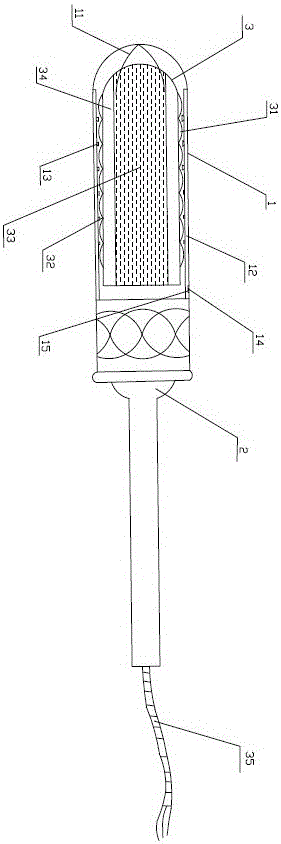
[0014] Combine now figure 1 The present invention is further described, a kind of analgesic applicator for intrauterine adhesiolysis, comprising an outer tube 1, an inner push rod 2 is arranged in the outer tube 1, and the inner push rod 2 and the outer tube 1 sliding fit, the front end of the inner push rod 2 is provided with a drug soaking part 3 that can be separated from the inner push rod 2, and the drug soaking part 3 is adsorbed with tetracaine glue, and the inner push rod 2 is The rod 2 is provided with a base, and the medicine soaking part 3 is located on the base. The front end of the overtube 1 is provided with an opening 11 , and the opening 11 at the front end of the overtube 1 is closed in the first state and pressed open by the drug soaking part 3 in the second state. The inner push rod 2 pushes the drug-soaking part 3 out from the opening 11 at the front end of the outer sleeve 1, and the inner wall of the outer sleeve 1 is provided with an inwardly protruding...


PUM


Abstract

The invention discloses an analgesia dosing device used after a tissue adhesion separation operation within the uterine-cavity. According to the injection dosing device used for postoperative analgesia, a medicine dipping part of the dosing device contains tetracaine gel, the medicine dipping part is left in the vagina after the operation, medicine can continuously anaesthetize the cervix uterus and pudendal nerves, and thus the effect of postoperative analgesia is achieved. The medicine dipping part can be taken out through a traction rope on the next day, no operative instruments are required, a dosing groove which protrudes inwards is formed in the inner wall of an outer sleeve, and atomization spraying heads are arranged in inward through holes of the dosing groove. In the using process, the needle tubing is inserted into a check valve of a medicine inlet in the outer wall of the outer sleeve for dosing and pressurization, atomized drugs can be evenly sprayed onto the surface of the medicine dipping part through the atomization spraying heads, the medicine can arrive at the inside of a human body after being adsorbed onto the surface of the medicine dipping part and performs slow and long-term release, and therefore the effect of postoperative analgesia is achieved.

Description

technical field [0001] The invention specifically relates to the technical field of medical instruments, in particular to an analgesic applicator for postoperative separation of intrauterine adhesions. Background technique [0002] Intrauterine adhesions are a group of diseases in which the uterine cavity is partially or completely occluded due to endometrial injury. With the development and wide application of hysteroscopic technology, especially the continuous improvement and promotion of hysteroscopic separation of intrauterine adhesions, intrauterine adhesions can be treated better. In recent years, indwelling balloon stents under hysteroscopy has become a widely used treatment for preventing postoperative uterine re-adhesion. However, during the operation, due to operations such as pressurizing the uterus, expanding the uterus, curettage, clamping, etc., and the compression of the uterine wall by the expansion balloon, the postoperative pain caused by the patient is un...

Claims


Application Information

Patent Timeline
Patent Type & Authority Applications(China)
IPC IPC(8): A61M31/00A61M19/00
CPCA61M19/00A61M31/00A61M2210/1475
Inventor 张丽青林凤施克俭徐旭仲
Owner THE FIRST AFFILIATED HOSPITAL OF WENZHOU MEDICAL UNIV
Why Eureka
Try Eureka