A self-image electrospinning scaffold and its preparation method

An electrospinning and self-imaging technology, which is applied in the field of medical devices, can solve the problems of non-degradable metal stents, inability to develop polymer stents, and inability to display the shape of stents well, so as to prevent thrombus formation

Active Publication Date: 2021-05-18
乐普(深圳)国际发展中心有限公司
View PDF12 Cites 0 Cited by
  • Summary
  • Abstract
  • Description
  • Claims
  • Application Information

AI Technical Summary

Problems solved by technology

[0002] Vascular stents are used to treat diseases such as vascular stenosis. Common vascular stents are mainly made of metal or polymer. Metal stents can be visualized under X-rays to facilitate detection of vascular stents, but metal stents are non-degradable and limit the normal pulsation of blood vessels; Degradable polymer stent overcomes this problem, but the polymer stent cannot be developed under X-ray. The existing development method is to add a development ring to the polymer stent or to coat the development layer, so that the polymer stent has the development function of
However, the developing ring can only show the positions of the two ends of the stent, and cannot show the shape of the stent very well, which brings certain difficulties to the later follow-up observation

Method used


Image

  • A self-image electrospinning scaffold and its preparation method
  • A self-image electrospinning scaffold and its preparation method

Examples


Embodiment 1

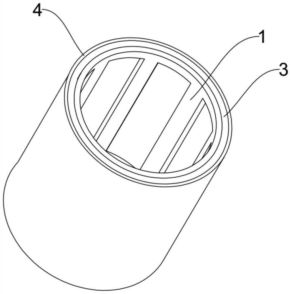
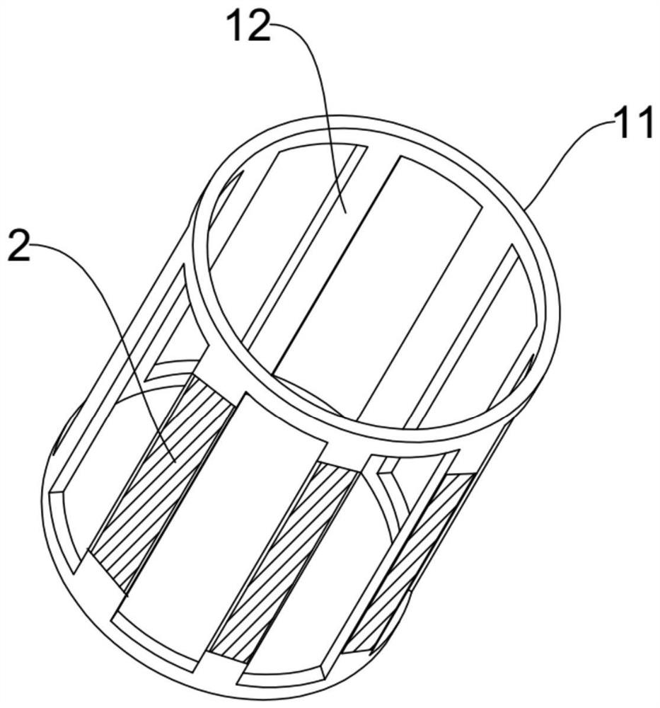
[0026] Embodiment 1: A self-developing electrospinning stent, comprising a stent body 1, a developing layer 2, a spinning layer 3 and a protective layer 4, the stent body 1 includes two symmetrically arranged support rings 11, between the two supports Four support bars 12 are arranged between the rings 11, and the two support rings 11 are coaxially arranged, and the two ends of the support bars 12 are respectively connected with the two support rings 11, as figure 2 As shown, a tubular shape is formed between the support ring 11 and the support bars 12 , and the support bars 12 are evenly distributed on the circumference of the support ring 11 . The developing layer 2 is arranged on the outer surface of the support bar 12, the spinning layer 3 is wrapped around the outer wall of the stent body 1, and the protective layer 4 is coated on the outside of the spinning layer 3 , the thickness of the protection layer 4 is 1um.

[0027] The method for preparing the stent of this emb...

Embodiment 2

[0033] Embodiment 2: A self-developing electrospinning stent, comprising a stent body 1, a developing layer 2, a spinning layer 3 and a protective layer 4, the stent body 1 includes two symmetrically arranged support rings 11, between the two supports Six support bars 12 are arranged between the rings 11, and the two support rings 11 are coaxially arranged, and the two ends of the support bars 12 are respectively connected with the two support rings 11, as figure 2 As shown, a tubular shape is formed between the support ring 11 and the support bars 12 , and the support bars 12 are evenly distributed on the circumference of the support ring 11 . The developing layer 2 is arranged on the outer surface of the support bar 12, the spinning layer 3 is wrapped around the outer wall of the stent body 1, and the protective layer 4 is coated on the outside of the spinning layer 3 , the thickness of the protective layer 4 is 2um.

[0034] The method for preparing the stent of this embo...

Embodiment 3

[0039] Embodiment 3: A self-developing electrospinning stent, including a stent body 1, a developing layer 2, a spinning layer 3 and a protective layer 4, the stent body 1 includes two symmetrically arranged support rings 11, between the two support rings Eight support bars 12 are arranged between the rings 11, and the two support rings 11 are coaxially arranged, and the two ends of the support bars 12 are respectively connected with the two support rings 11, as figure 2 As shown, a tubular shape is formed between the support ring 11 and the support bars 12 , and the support bars 12 are evenly distributed on the circumference of the support ring 11 . The developing layer 2 is arranged on the outer surface of the support bar 12, the spinning layer 3 is wrapped around the outer wall of the stent body 1, and the protective layer 4 is coated on the outside of the spinning layer 3 , the thickness of the protection layer 4 is 3um.

[0040] The method for preparing the stent of thi...


PUM

PropertyMeasurementUnit
molecular weightaaaaaaaaaa

Abstract

The invention discloses a self-developing electrospinning stent, which comprises a stent body, a developing layer, a spinning layer and a protective layer. The stent body includes two symmetrically arranged support rings and a connecting ring between the two The support bar, the developing layer is arranged on the surface of the support bar, the spinning layer is wrapped around the outer wall of the stent body, and the drug-loaded layer is coated on the outside of the spinning layer. The preparation method is as follows: (a) laser engraving the pipe material, (b) preparing a developing mixed solution to obtain a developing layer; (c) spraying a spinning layer on the outer wall of the stent body; (d) spraying by ultrasonic atomization to obtain a protective layer, dry. The spinning layer in the stent of the present invention provides a radial support force for the stent, so that the stent always has a certain strength of support force. The treatment efficiency of the stent is improved.

Description

technical field [0001] The invention belongs to the technical field of medical devices, and in particular relates to a self-image electrospinning support and a preparation method thereof. Background technique [0002] Vascular stents are used to treat diseases such as vascular stenosis. Common vascular stents are mainly made of metal or polymer. Metal stents can be visualized under X-rays to facilitate detection of vascular stents, but metal stents are non-degradable and limit the normal pulsation of blood vessels; Degradable polymer stent overcomes this problem, but the polymer stent cannot be developed under X-ray. The existing development method is to add a development ring to the polymer stent or to coat the development layer, so that the polymer stent has the development function. However, the developing ring can only show the positions of the two ends of the stent, and cannot show the shape of the stent very well, which brings certain difficulties to the later follow-...

Claims


Application Information

Patent Timeline
Patent Type & Authority Patents(China)
IPC IPC(8): A61F2/82A61L31/10A61L31/14A61L31/16A61L31/18
CPCA61F2/82A61F2002/823A61F2250/0098A61L31/10A61L31/146A61L31/148A61L31/16A61L31/18A61L2300/42A61L2300/604C08L67/04C08L89/00
Inventor 万明洲王刚王亮陈果
Owner 乐普(深圳)国际发展中心有限公司
Why Eureka
Try Eureka