Whole eyeball cornea preservation method

A preservation method and eyeball technology, which is applied in the field of medical ophthalmology, can solve the problems of high price, short preservation time, and no activity of corneal endothelium, and achieve the effect of maintaining activity, avoiding toxic and side effects, and prolonging the storage time in wet room

Inactive Publication Date: 2019-10-29
李冰
View PDF7 Cites 1 Cited by
  • Summary
  • Abstract
  • Description
  • Claims
  • Application Information

AI Technical Summary

Problems solved by technology

[0012] The present invention aims at the deficiencies of the above-mentioned prior art, provides a kind of not only simple but also can prolong the preservation method of cornea of ​​whole eyeball cornea, solves several methods in the prior art that preservation time is short and expensive, and there is no activity of corneal endothelium question

Method used


Image

  • Whole eyeball cornea preservation method
  • Whole eyeball cornea preservation method
  • Whole eyeball cornea preservation method

Examples


example 1

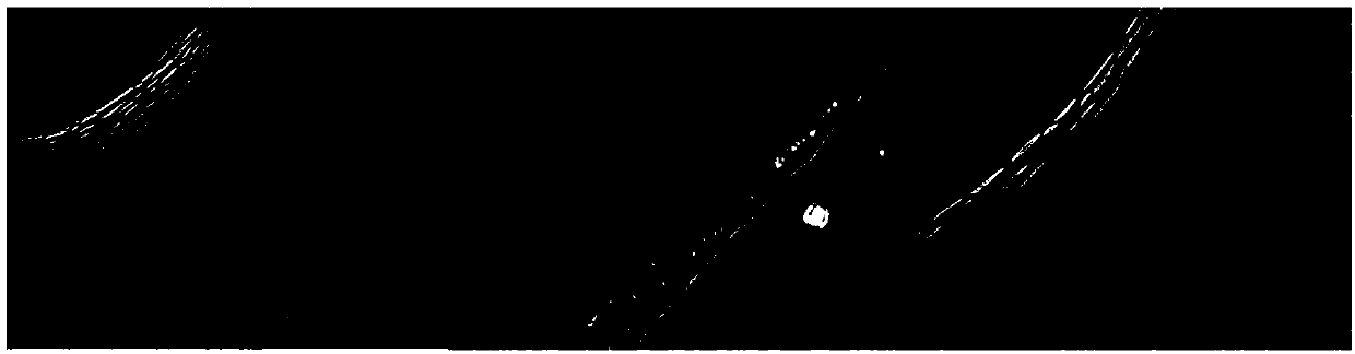
[0087] Example 1. Patient A is a keratoconus patient. The cornea preserved for 4 days by the preservation method of the present invention was operated on. The cornea was transparent three months after the operation, without obvious rejection or edema. The comparison chart before and after operation is attached Figure 18 shown. The analysis chart of corneal endothelial count at 3 months after keratoplasty Figure 19 As shown, the morphology of corneal endothelial cells can be seen to be normal.

example 2

[0088] Example 2. Patient B is a patient with leukoplakia. The cornea preserved for 3 days by this preservation method was used for surgery. The cornea was transparent 6 months after the operation, without obvious rejection or edema. The comparison chart before and after operation is attached Figure 20 shown. The analysis chart of corneal endothelial count in the patient's eye 6 months after keratoplasty Figure 21 As shown, it can be seen that the morphology of corneal endothelial cells is normal, and the density of endothelial cells is acceptable.

example 3

[0089] Example 3. Patient C is a patient with corneal dystrophy. The cornea was preserved for 2 days using this preservation method for surgery. The cornea was transparent 3 months after the operation, without obvious rejection or edema. The comparison chart before and after operation is attached Figure 22 shown. The analysis chart of corneal endothelial count of the patients 3 months after keratoplasty Figure 23 As shown, the corneal endothelial cells can be seen to be normal in shape and smooth.


PUM


Abstract

The invention discloses a whole eyeball cornea preservation method. The method utilizes a sodium chondroitin sulfate-hyaluronate viscoelastic agent for carrying out eyeball preservation to prolong cornea preservation time, and is an improved method for preserving a cornea in a moist chamber. Specifically the cornea preservation method comprises the following steps: eyeball treatment: taking down an eyeball within 2 h after a donor dies, performing disinfection treatment as quickly as possible and then evacuating aqueous humor at the same time, and injecting a chondroitin sulfate-sodium hyaluronate solution into anterior chamber, wherein the chondroitin sulfate-sodium hyaluronate solution is a colorless transparent gelatinous solution consisting of 3% sodium hyaluronate, 4% sodium chondroitin sulfate and a physiological buffer balance salt; and placing the whole eyeball after treatment in a sterile tank, and placing the sterile tank in a 4 DEG C moist chamber environment for preservation. A cornea preservation time can be extended to 4 d by using the method, activity of corneal endothelial cells is not significantly decreased, and a moist chamber preservation time is greatly increased.

Description

technical field [0001] The invention relates to the technical field of medical ophthalmology, in particular to a method for preserving the cornea of ​​the whole eyeball which is simple and can prolong the preservation time of the cornea. Background technique [0002] The cornea is a transparent film at the front of the eye. When the cornea is damaged or diseased, it needs to be treated with a corneal transplant. The cornea used in corneal transplantation can be an artificial biological cornea or a human donated cornea. [0003] Human donated corneas need to be processed and preserved before corneal transplantation. At present, the preservation methods of the cornea donated by the human body in the prior art include: [0004] Wet room storage: For wet room storage, the entire donor eye is sterilized and placed in a sterile jar and placed in a suitable humid environment. If the time between the death of the donor and enucleation is shorter than 4~6h, the donor eye can gene...

Claims


Application Information

Patent Timeline
Patent Type & Authority Applications(China)
IPC IPC(8): A01N1/02
CPCA01N1/0226
Inventor 盛敏杰李冰陈丽
Owner 李冰
Why Eureka
Try Eureka