Image processing apparatus, image processing system and image processing method

a technology of image processing apparatus and image processing system, which is applied in the direction of static indicating devices, instruments, cathode-ray tube indicators, etc., can solve the problem that the presentation of quantitative information on some of the indices as in the above conventional art cannot be said to be high in versatility, and the operation is a large load on the pathologist, so as to achieve the effect of supporting the diagnosis operation

Active Publication Date: 2022-01-04
DAINIPPON SCREEN MTG CO LTD
View PDF5 Cites 0 Cited by
  • Summary
  • Abstract
  • Description
  • Claims
  • Application Information

AI Technical Summary

Benefits of technology

[0010]This invention was developed in view of the above problem and an object thereof is to provide a technique capable of effectively supporting a diagnosis operation by a doctor by presenting various pieces of information obtained from an image easily and in a multifaceted way in displaying the image captured by imaging a pathological specimen.
[0016]Further, the “visual information” is various pieces of information which enables the user to easily visually recognize a specific region of the source image being the region extracted as the pathological finding and properties and the like of the pathological finding in this region. Modes of the visual information possibly include, for example, color coding, the emphasis of the contour or shading of the region, the attachment of markers by symbols or graphics or character information. These may be combined as appropriate.
[0017]In the inventions thus configured, the information on the pathological findings required by the user (doctor who diagnoses) can be easily presented according to a request and effectively support a diagnosis operation. The reason for that is as follows. At a pathological diagnosis site, a pathological specimen is observed from various perspectives and comprehensively diagnosed. Thus, a support method for displaying and presenting various pathological findings extracted from the pathological specimen in various display modes is more desirable than the presentation of only a quantitative evaluation result on a specific pathological finding.
[0024]As described above, the invention can be realized to cover various display modes required at a pathological diagnosis site. Further, in each display mode, an image can be variously changed according to a request of a user (doctor). Thus, various pieces of information obtained from the image can be presented in a mode easily understandable by the user and in a multifaceted way, and a diagnosis operation by the doctor can be effectively supported.

Problems solved by technology

Such an operation is a large load on the pathologist and possibly causes a variation in diagnosis result.
However, at the moment, it has not yet reached to automate an entire operation performed by a pathologist for diagnosis, and judgment based on the pathologist's experiences remains to be necessary.
Thus, the presentation of quantitative information on some of indices as in the above conventional art cannot be necessarily said to be high in versatility.
This staining method can separately stain cytoplasms and cell nuclei, but is not for selectively staining specific cell species, disease sites and the like.
Specifically, many of conventional techniques focus on features unique to specific cells and diseases and utilize these features, and a range of its application has been restrictive.

Method used


Image

  • Image processing apparatus, image processing system and image processing method
  • Image processing apparatus, image processing system and image processing method
  • Image processing apparatus, image processing system and image processing method

Examples


Embodiment Construction

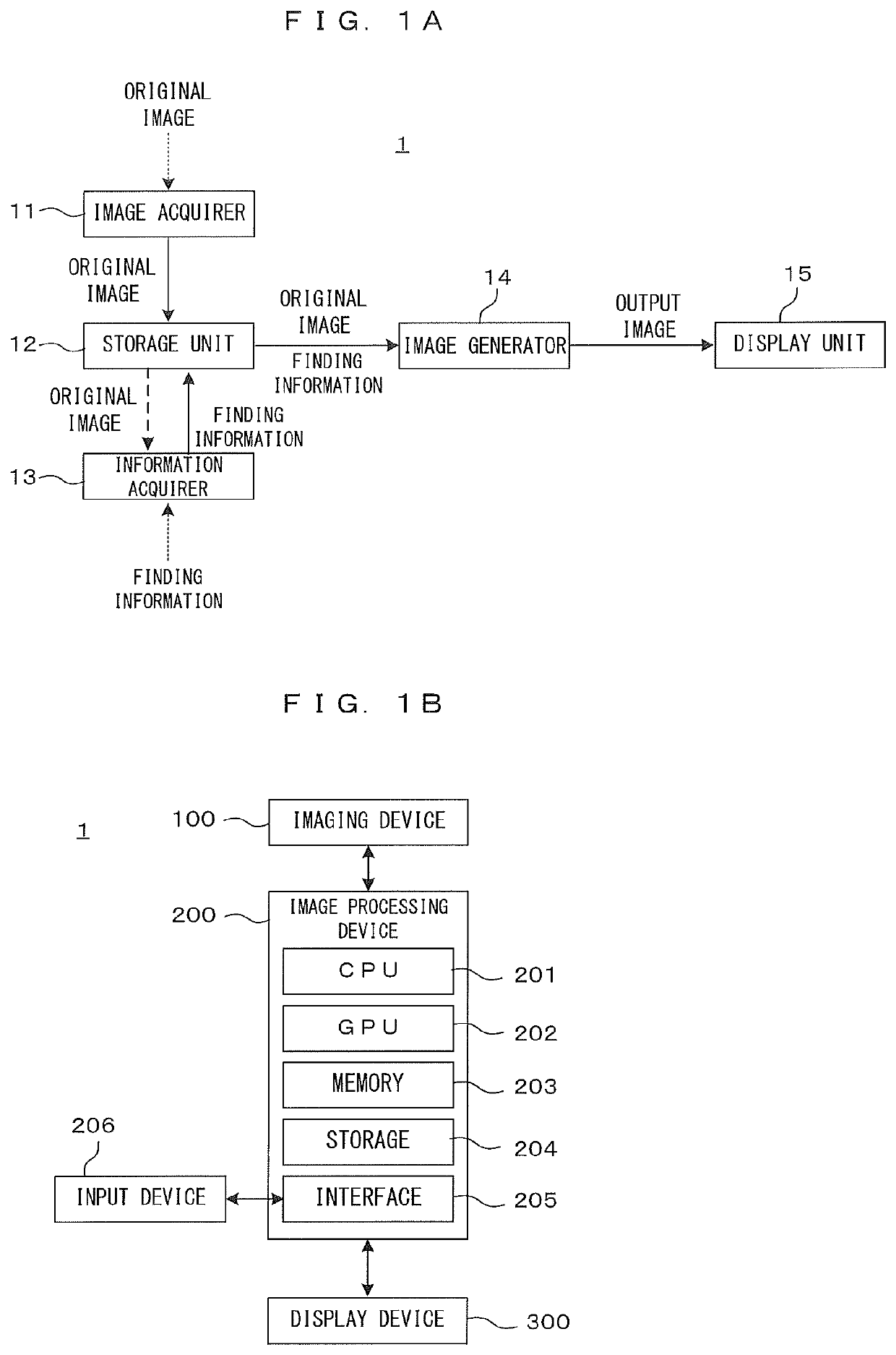
[0034]FIGS. 1A and 1B are diagrams showing a schematic configuration of one embodiment of an image processing system according to the invention. More specifically, FIG. 1A is a block diagram conceptually showing functional blocks which should be included in the image processing system 1 to carry out the invention. Further, FIG. 1B is a block diagram showing a more specific hardware configuration. This image processing system 1 is a system for supporting an operation of a user (specifically, a pathologist), who observes and diagnoses a pathological specimen collected from a patient or a subject, from the aspect of an image processing.

[0035]This image processing system 1 can be applied to pathologic diagnoses for various diseases in various organs, and application targets thereof are not particularly limited. However, if it is necessary to mention a particularly specific case example in the following description, a pathological diagnosis of a brain tumor based on an image of a patholo...


PUM


Abstract

An image processing apparatus includes an image acquirer for obtaining an original image of a pathological specimen, an information acquirer for obtaining finding information relating to pathological finding and an image generator for generating an output image including a processed image obtained by superimposing visual information based on the finding information on a source image. A relative density between the source image and the visual information is set according to an operation input from a user. Processing modes for generating the output image include a first mode for generating the output image in which the source image and the processed image are arranged on one screen and a second mode for generating the output image in which a wide range image at a relatively low magnification and an enlarged image representing a partial region in the wide range image at a higher magnification on one screen.

Description

CROSS REFERENCE TO RELATED APPLICATION[0001]The disclosure of Japanese Patent Application No. 2020-012483 filed on Jan. 29, 2020 including specification, drawings and claims is incorporated herein by reference in its entirety.BACKGROUND OF THE INVENTION1. Field of the Invention[0002]This invention relates to an image processing technique for processing an original image captured by imaging a pathological specimen. Particularly, this invention relates to a technique for supporting a diagnosis by a pathologist by reflecting and presenting pathological findings extracted by an image analysis on a displayed image.2. Description of the Related Art[0003]At a pathological diagnosis site, a skilled doctor (pathologist) has observed a pathological specimen collected from a patient and diagnosed by comprehensively judging a state of the pathological specimen. Such an operation is a large load on the pathologist and possibly causes a variation in diagnosis result. Thus, the development of a te...

Claims


Application Information

Patent Timeline
Patent Type & Authority Patents(United States)
IPC IPC(8): G09G5/00G09G5/36
CPCG09G5/363G09G2340/12G09G2380/08G09G2340/045G09G2354/00
Inventor OGI, HIROSHIMORIWAKI, SANZOFURUTA, TOMOYASU
Owner DAINIPPON SCREEN MTG CO LTD
Why Eureka
Try Eureka