Three dimensional atrium-ventricle plane detection

ultrasound technology, applied in the field of ultrasound system for detecting a three-dimensional atrium ventricle plane, can solve the problems of difficult or impossible direct assessment of new parameters, unacceptable inter-observer variability, and hampered evaluation of cardiac function

Inactive Publication Date: 2006-03-16
GENERAL ELECTRIC CO
View PDF8 Cites 51 Cited by
  • Summary
  • Abstract
  • Description
  • Claims
  • Application Information

AI Technical Summary

Benefits of technology

[0017] One embodiment of the present invention relates to a system and method for generating an image responsive to moving cardiac structure and blood. One or more embodiments of the present invention relates to a an ultrasound machine adapted to generate an image responsive to moving cardiac structure and blood. This embodiment of the method comprise acquiring 3D ultrasound data containing at least one view of the moving cardiac structure and blood and identifying an AV-plane using the at least one acquired view. The method further comprises generating a cardiac 3D image using at least the identified AV-plane.

Problems solved by technology

Evaluation of cardiac function has been hampered by a lack of well-established parameters that may be used to increase the accuracy and objectivity in the assessment of diseases, coronary artery diseases for example.
It has been shown that inter-observer variability between echo-centers is unacceptably high due to the subjective nature of the cardiac motion assessment.
Many of the new parameters have been difficult or impossible to assess directly by visual inspection of the ultrasound images generated in real-time.
The quantification has typically required a post-processing step with tedious, manual analysis to extract the necessary parameters.
Time intensive post-processing techniques or complex, computation-intensive real-time techniques are undesirable.

Method used


Image

  • Three dimensional atrium-ventricle plane detection
  • Three dimensional atrium-ventricle plane detection
  • Three dimensional atrium-ventricle plane detection

Examples


Embodiment Construction

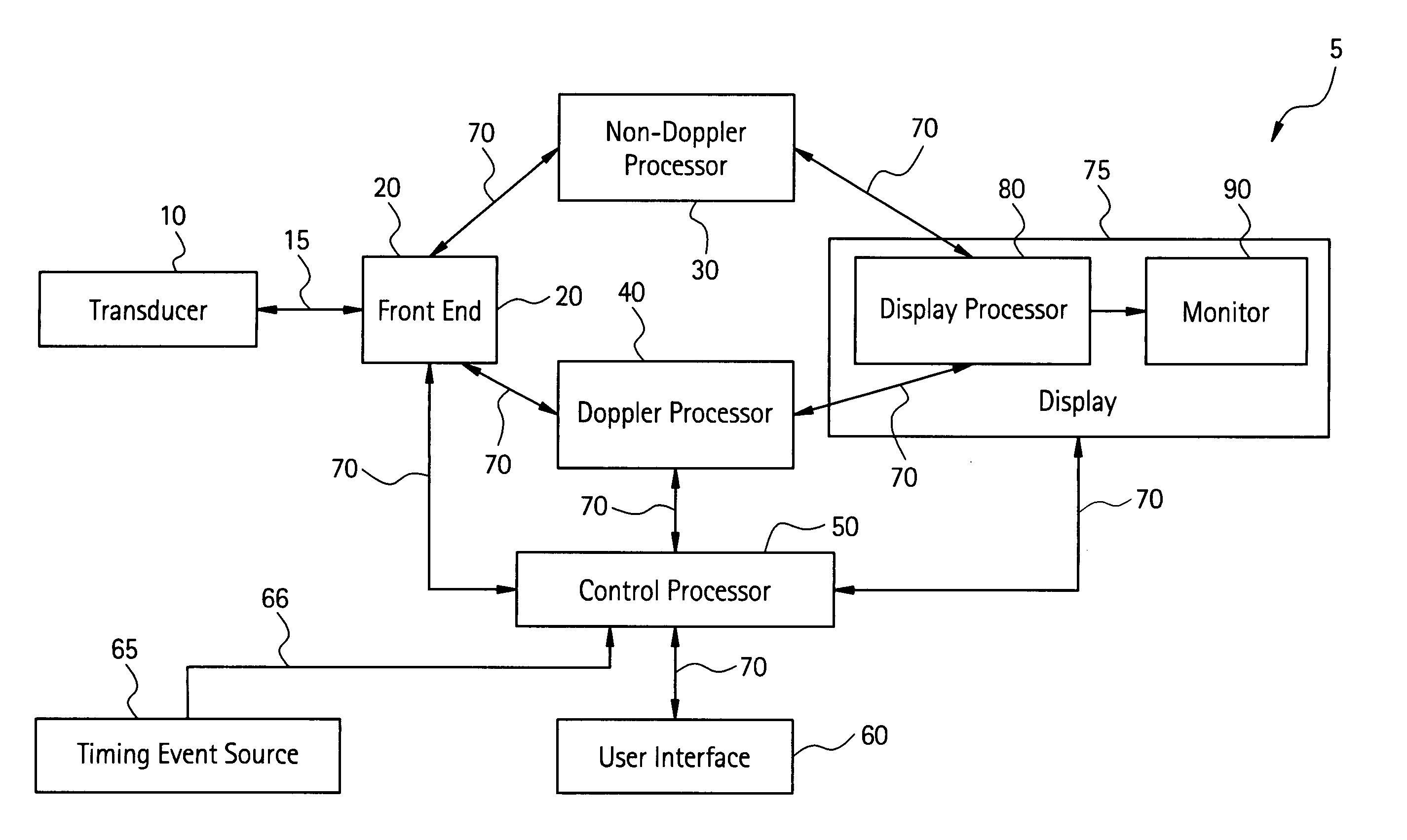
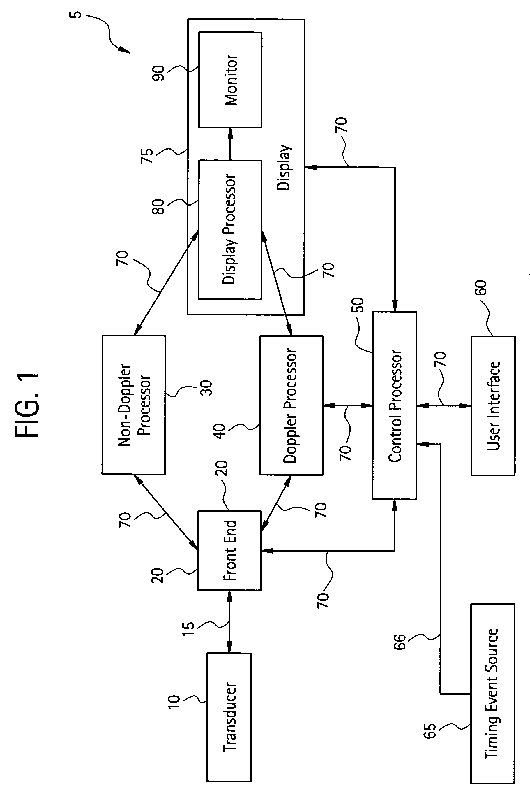
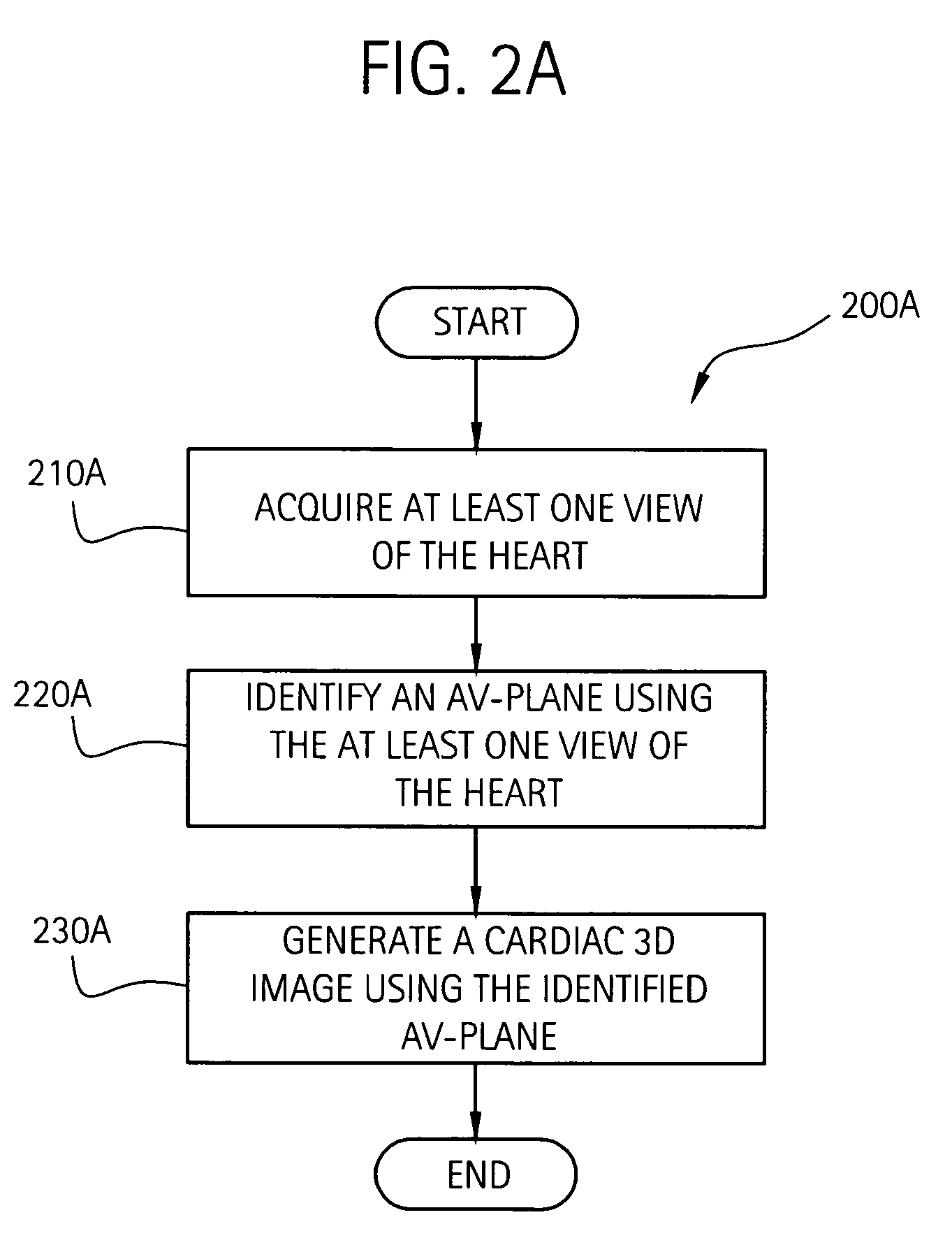
[0037] An embodiment of the present invention relates to an ultrasound system for detecting a 3D AV-plane. More specifically, an embodiment of the present invention relates to an ultrasound system for imaging a heart, identifying at least an AV-plane of the heart and forming a cardiac three-dimensional 3D image of at least a portion of the heart using at least the AV-plane. Moving cardiac structure is monitored to accomplish this function. As used herein, the term structure comprises non-liquid and non-gas matter, such as cardiac tissue for example. An embodiment of the present invention provides improved, real-time visualization and quantative assessment of certain clinically relevant or key parameters of the heart. The moving structure is characterized by a set of analytic or key parameter values corresponding to anatomical points within a myocardial segment of the heart. The set of analytic or key parameter values may comprise, for example, tissue velocity values, time-integrated...


PUM


Abstract

The present invention relates to a method and apparatus for generating at least a 3D image responsive to moving cardiac structure and blood, and extracting clinically relevant information based on anatomical landmarks located within the heart. One embodiment of the present invention comprises at least a front end and at least one processor. The front-end is arranged to transmit ultrasound waves into the moving cardiac structure and blood of a heart and generate received signals in response to ultrasound waves backscattered from the said moving cardiac structure and blood. The at least one processor responsive to the received signals to acquire 3D ultrasound data containing at least one view of the heart, identify an AV-plane using the at least one acquired view, and generate a cardiac 3D image of at least a portion of the heart using at least one identified AV-plane. At least the 3D image may be displayed to a user.

Description

RELATED APPLICATIONS / INCORPORATION BY REFERENCE [0001] This application is related to, and claims benefit of and priority from, Provisional Application No. 60 / 606,041, filed Aug. 31, 2004, titled “THREE DIMENSIONAL ATRIUM-VENTRICLE PLANE DETECTION”, the complete subject matter of which is incorporated herein by reference in its entirety. [0002] complete subject matter of each of the following U.S. patent applications is incorporated by reference herein in their entirety: [0003] U.S. patent application Ser. No. 10 / 248,090 filed on Dec. 17, 2002. [0004] U.S. patent application Ser. No. 10 / 064,032 filed on Jun. 4, 2002. [0005] U.S. patent application Ser. No. 10 / 064,083 filed on Jun. 10, 2002. [0006] U.S. patent application Ser. No. 10 / 064,033 filed on Jun. 4, 2002. [0007] U.S. patent application Ser. No. 10 / 064,084 filed on Jun. 10, 2002. [0008] U.S. patent application Ser. No. 10 / 064,085 filed on Jun. 10, 2002. [0009] U.S. Provisional Patent Application Ser. No. 60,605,939 (Attorney ...

Claims


Application Information

Patent Timeline
Patent Type & Authority Applications(United States)
IPC IPC(8): A61B8/02
CPCA61B8/08A61B8/483A61B8/0883
Inventor OLSTAD, BJORN
Owner GENERAL ELECTRIC CO
Why Eureka
Try Eureka