Radiation tomographic imaging apparatus and radiation tomographic imaging method

a tomographic imaging and radiation tomography technology, applied in tomography, applications, instruments, etc., can solve the problems of unsatisfactory image quality and unsatisfactory heartbeat phase, and achieve the effect of reducing the radiation exposure dose of the subject, improving the efficiency of diagnosis and improving operability

Inactive Publication Date: 2007-06-28
GE MEDICAL SYST GLOBAL TECH CO LLC
View PDF7 Cites 6 Cited by
  • Summary
  • Abstract
  • Description
  • Claims
  • Application Information

AI Technical Summary

Benefits of technology

[0027] The radiation tomographic imaging apparatus or method in accordance with the present invention allows the increase of efficiency of the diagnosis of a subject because of the improved operability at the time when confirming a t

Problems solved by technology

However, when the heartbeat is tachycardiac if subject is tense by holding the breath or when in arr

Method used


Image

  • Radiation tomographic imaging apparatus and radiation tomographic imaging method
  • Radiation tomographic imaging apparatus and radiation tomographic imaging method
  • Radiation tomographic imaging apparatus and radiation tomographic imaging method

Examples


Embodiment Construction

[0032] [Overview of an X-Ray CT Apparatus]

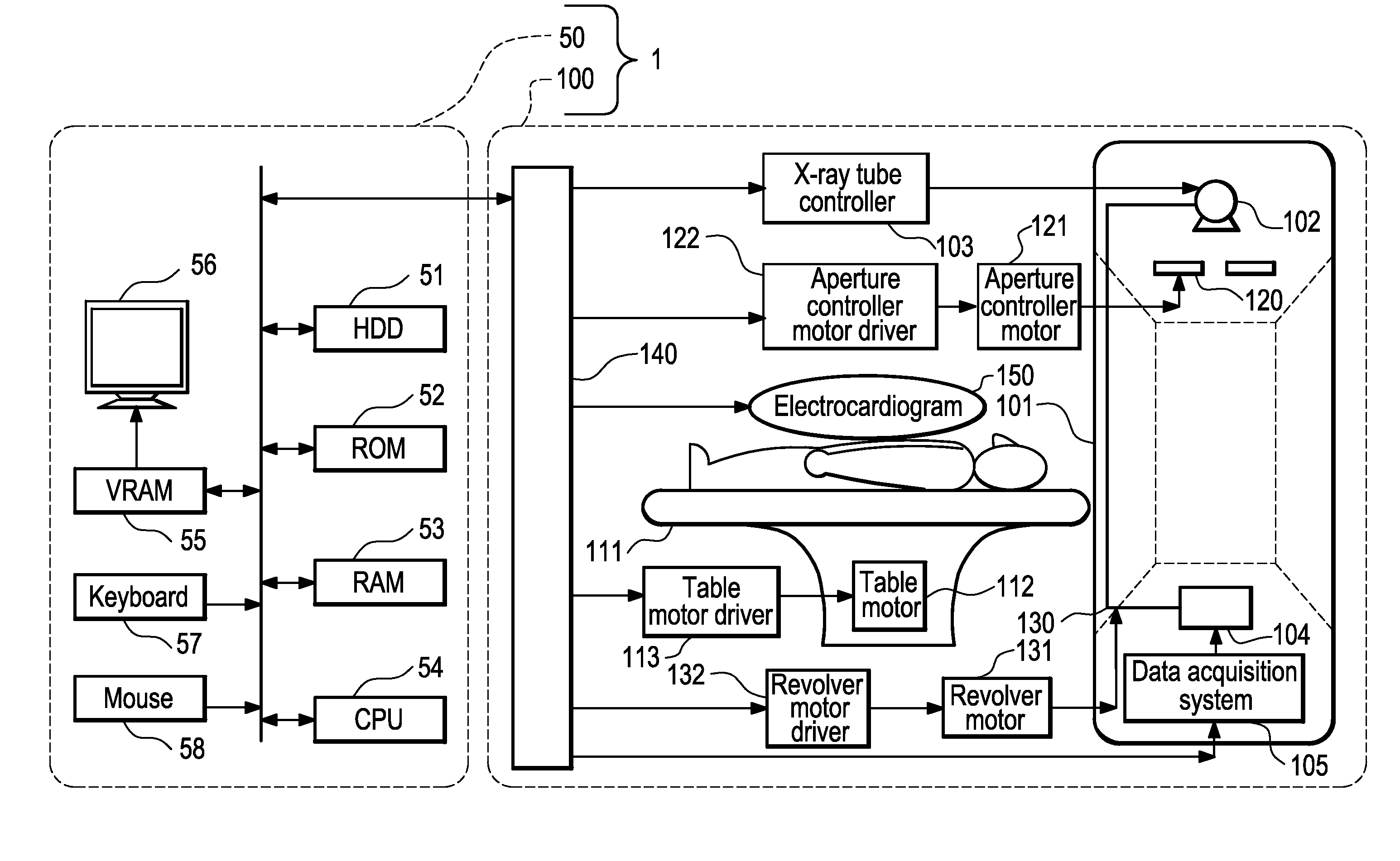
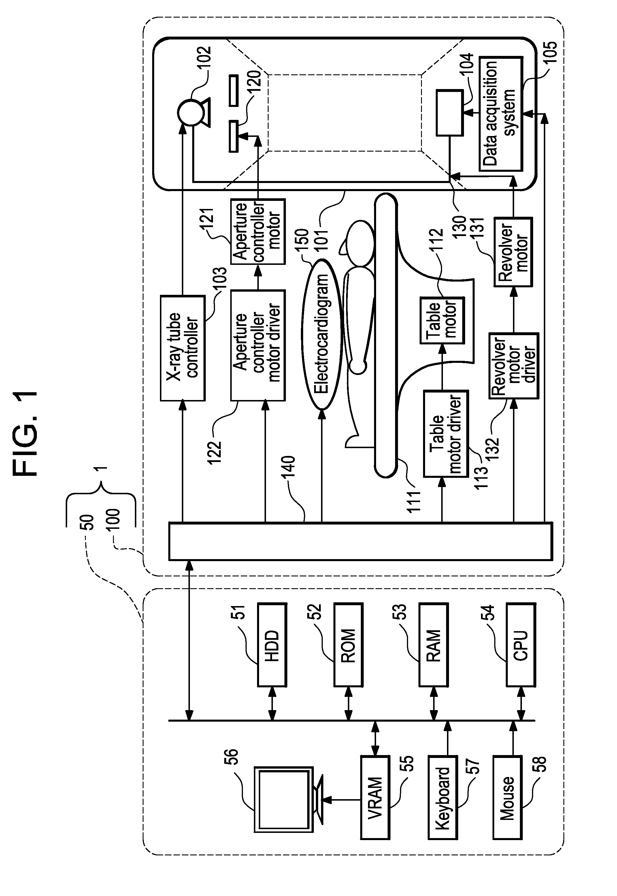
[0033] Now referring to FIG. 1, there is shown an overview of an X-ray CT apparatus 1 in accordance with the preferred embodiment. As shown in the figure, the apparatus includes a gantry 100 for emitting X-ray to the subject and for detecting X-ray transmitted through the subject, and an operation console 50 for reconstructing an X-ray tomographic image based on the data transferred from the gantry 100 and for outputting and displaying.

[0034] The gantry 100 includes a CT controller unit 140 for managing the entity and is connected to a variety of equipment as will be described below.

[0035] Inside the gantry 100, there are provided an X-ray tube 102 which is the source of X-ray, an X-ray tube controller 103 connected to the X-ray tube 102, a collimator 120 having an aperture for limiting the radiation range of the X-ray, an aperture controller motor 121 for adjusting the aperture width of the collimator 120, and an aperture controller motor...


PUM


Abstract

There is provided a radiation tomographic imaging apparatus and a radiation tomographic imaging method which allows the ECG information and the X-ray tube current value to be monitored while obtaining required projection data for the image reconstruction. The radiation tomographic imaging method for forming a tomographic image of a subject by means of the radiation from the radiation source comprises the ECG wave output step for measuring the heartbeat of the heart of the subject to output as the ECG wave signal; a variable output step for varying the radiation output based on the ECG wave signal; a determining step for determining the tomographic image reconstructed based on the projection data obtained by the radiation output having been varied is good or no good; and a displaying step for displaying the ECG wave signal, the radiation output, and the reconstruction data area for forming a tomographic image if the image is no good.

Description

CROSS REFERENCE TO RELATED APPLICATIONS [0001] This application claims the benefit of Japanese Application No. 2005-376790 filed Dec. 28, 2005. BACKGROUND OF THE INVENTION [0002] The present invention relates to a radiation tomographic imaging method for performing a tomographic imaging of a subject by emitting radiation from the circumference of the subject, namely the patient to obtain data and to process the data. More specifically the present invention relates to the reconstruction of the image of a heart area. [0003] As the diagnostic apparatus for a lesion in a subject, a radiation tomographic imaging apparatus, such as an X-ray CT apparatus, for obtaining a tomographic image of a subject is widely used for the diagnosis. The X-ray CT apparatus is also widely used in the imaging of the heart area. [0004] When imaging the heart area, because the heart is beating all the time, the patient has an electrocardiogram attached thereon to monitor the function state of the heart such a...

Claims


Application Information

Patent Timeline
IPC IPC(8): A61B6/00G01N23/00G21K1/12H05G1/60
CPCA61B5/0402A61B6/032A61B6/463A61B6/488A61B6/503A61B6/541A61B5/318A61B6/03
Inventor SETO, MASARU
Owner GE MEDICAL SYST GLOBAL TECH CO LLC
Why Eureka
Try Eureka