Method and System for Intra Luminal Thrombus Detection

a luminal thrombus and detection method technology, applied in the field of methods and systems for intra luminal thrombus detection, can solve problems such as thrombosis formation, disruption or ruptur

Active Publication Date: 2008-07-03
INFRAREDX INC
View PDF13 Cites 15 Cited by
  • Summary
  • Abstract
  • Description
  • Claims
  • Application Information

AI Technical Summary

Benefits of technology

[0014]The above and other features of the invention including various novel details of construction and combinations of parts, and other advantages, will now be more particularly described with reference to the accompanying drawings and pointed out in the claims. It will be understood

Problems solved by technology

Another potential problem associated with vessels is the formation of thrombus.
In fact, disruption or rupture of atherosclerotic plaques appears to be the major cause of heart attacks and strokes, because, after the plaques r

Method used


Image

  • Method and System for Intra Luminal Thrombus Detection
  • Method and System for Intra Luminal Thrombus Detection
  • Method and System for Intra Luminal Thrombus Detection

Examples


Embodiment Construction

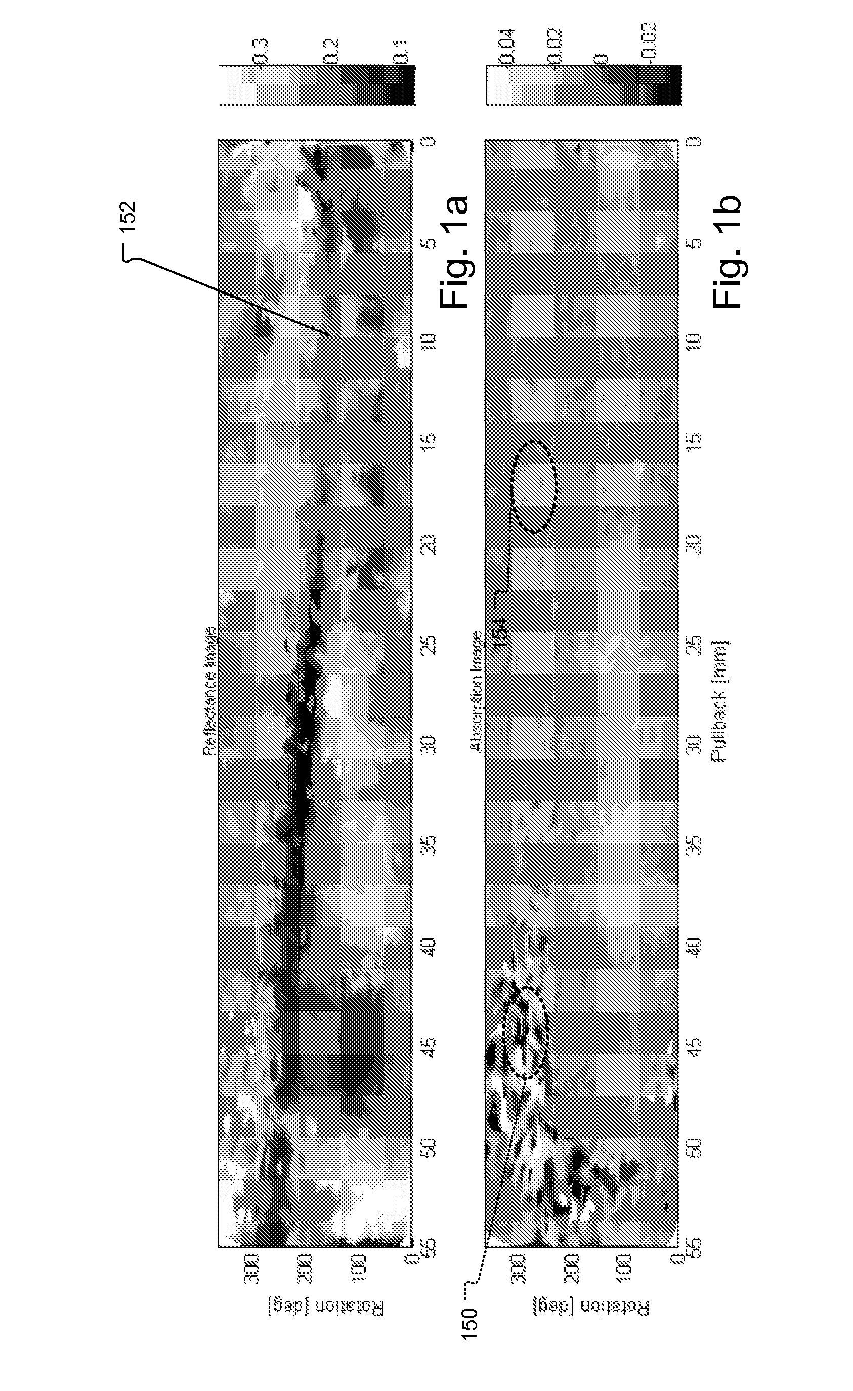
[0023]FIGS. 1a and 1b show a reflectance image and an absorption image generated by the near infrared (NIR) scanning of the inside of a blood vessel. These spectral measurements were collected through flowing blood.

[0024]The guide wire 152 can be seen as a dark shadow across the mean reflectance pullback image in FIG. 1a. For this image, an average of the reflectance spectrum across the full wavelength range is taken for each pixel. Thus, each pixel in the image is the average intensity of the reflectance spectrum at that point.

[0025]FIG. 1b is notable because it shows a distinct mottled pattern in regions including region 150 in the peak absorbance image at a pull back distance of approximately 40-55 millimeters corresponding to the location of an obstruction, such as a clot or thrombus. The absorption image is referred to as “peak absorbance” because here the average of each pixel is taken across a limited wavelength range (1200-1240 nm) where lipids have a strong absorbance signa...


PUM


Abstract

A method for detecting a complete or partial obstruction in a vessel through an intervening fluid includes receiving spectroscopic responses at different locations of a vessel wall, e.g., a vein or artery, through an intervening fluid, preferably blood. Spectroscopic responses are generated by irradiating the vessel wall at different locations and detecting spectra at those locations. In preferred embodiments, the radiation used is in the near infrared (NIR) region of the electromagnetic spectrum. The thrombus is located by determining whether fluctuations, spatial and/or spectral, of the spectral responses are indicative of thrombus.

Description

BACKGROUND OF THE INVENTION[0001]Intra luminal spectroscopic analysis devices have been developed and commercialized. One application concerns the assessment of the state of blood vessel walls such as required in the diagnosis of atherosclerosis. This is an arterial disorder involving the intimae of medium- or large-sized arteries, including the aortic, carotid, coronary, and cerebral arteries. Atherosclerotic lesions or plaques can contain complex tissue matrices, including collagen, elastin, proteoglycans, and extracellular and intracellular lipids with foamy macrophages and smooth muscle cells. In addition, inflammatory cellular components (e.g., T lymphocytes, macrophages, and some basophiles) can also be found in these plaques.[0002]The intra luminal spectroscopic analysis devices are used to extract information concerning the state of the vessels walls from the generated spectral information. Mathematical and statistical manipulations such as linear and non-linear regressions ...

Claims


Application Information

Patent Timeline
IPC IPC(8): A61B5/1455
CPCA61B5/0075A61B5/0086G01N21/359G01J3/42A61B5/02007
Inventor SUM, STEPHEN THOMASLISAUSKAS, JENNIFER BLANCHEMEESE, THOMAS M.
Owner INFRAREDX INC
Why Eureka
Try Eureka