Segmenting an angiography using an existing three-dimensional reconstruction

a three-dimensional reconstruction and segmenting technology, applied in the field of segmenting a two-dimensional angiography, can solve the problems of inaccurate geometric representation of stenosis geometry, low spatial resolution, and estimation of blood through vascular cross-sections, and achieve the effect of increasing the accuracy of three-dimensional angiography and facilitating compensation or correction

Active Publication Date: 2018-03-01
SIEMENS HEALTHCARE GMBH
View PDF1 Cites 11 Cited by
  • Summary
  • Abstract
  • Description
  • Claims
  • Application Information

AI Technical Summary

Benefits of technology

[0028]This is advantageous in that the individual angiographic recordings may profit from spatial information about the three-dimensional reconstruction and may be improved, and the two angiographic recordings may be improved in relation to one another (e.g., registered more accurately with one another). The accuracy of the three-dimensional angiography is thus increased. The method may also be carried out automatically or fully automatically.
[0029]In a further embodiment, a three-dimensional angiography of the vessel of the body may be generated from the two or mo...

Problems solved by technology

The disadvantage is the comparably minimal spatial resolution and thus an inaccurate geometrical representation of the stenosis geometry.
The disadvantage is the estimation of the blood through the vascular cross-sections.
Small errors may already have significant repercussions.
Estimating the blood flow via the contrast agent dynamics with the angio-FFR method is also complicated and difficult.
It is also disadvantageous that the angio-FFR method provides no information relating to a state of the myocardial mass, which is important, for example, in order t...

Method used


Image

  • Segmenting an angiography using an existing three-dimensional reconstruction
  • Segmenting an angiography using an existing three-dimensional reconstruction

Examples


Embodiment Construction

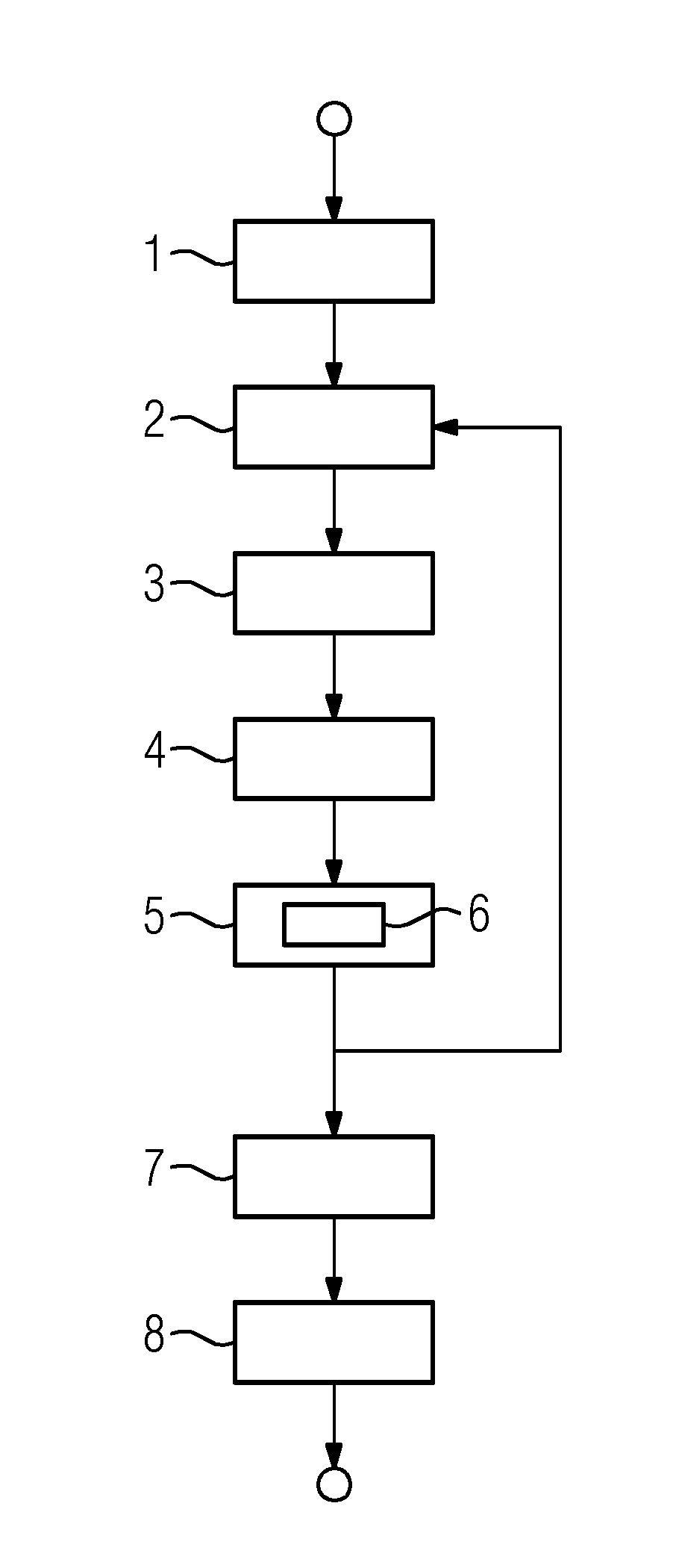
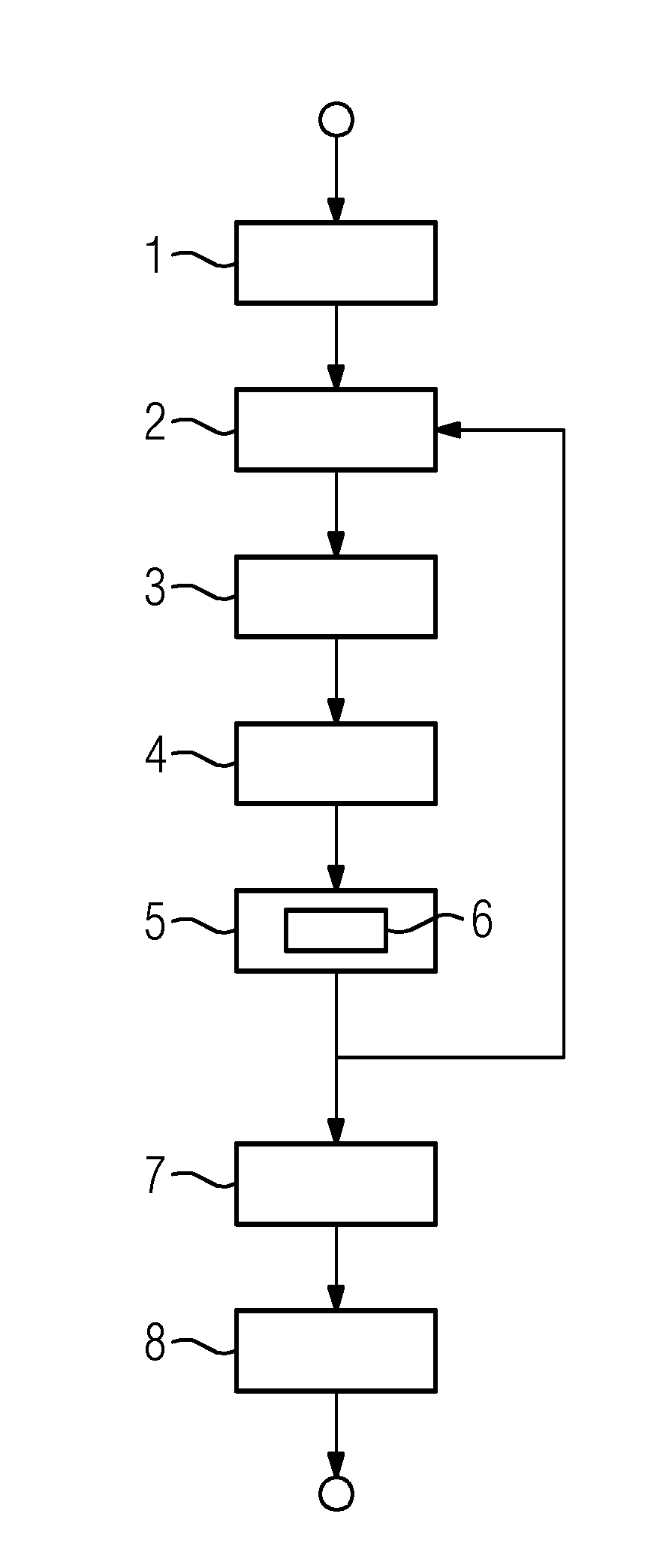
[0034]In a first act 1, a three-dimensional reconstruction of a vessel of a body (e.g., a coronary blood vessel) is provided. A three-dimensional reconstruction is provided in the form of a recorded data record. The recorded data record is produced by a first imaging device (e.g., a computed tomography (CT) device). A next act 2 involves further providing a first two-dimensional angiographic recording of the coronary blood vessel of a further imaging device (e.g., an x-ray device). This further provision 2 also takes place on the computing apparatus (e.g., in the form of an angiography data record).

[0035]In the example shown, the recorded data record and thus the three-dimensional reconstruction of the vessel of the body are thus registered 3 with the angiography data record, and thus, the two-dimensional recording of the vessel of the body that is present, for example, as a coronary blood vessel. This is followed by projecting 4 spatial information of the three-dimensional reconstr...


PUM


Abstract

A method for segmenting a two-dimensional angiographic recording of a vessel of a body using a computing apparatus includes providing a three-dimensional reconstruction of the vessel of the body to the computing apparatus. The two-dimensional angiographic recording of the vessel of the body is provided on the computing apparatus. The three-dimensional reconstruction of the vessel of the body is registered with the two-dimensional recording of the vessel of the body. Spatial information of the three-dimensional reconstruction is projected onto the two-dimensional recording, and the two-dimensional recording is segmented using the spatial information projected onto the two-dimensional recording.

Description

[0001]This application claims the benefit of DE 10 2016 215 971.2, filed on Aug. 25, 2016, which is hereby incorporated by reference in its entirety.BACKGROUND[0002]The present embodiments relate to segmenting a two-dimensional angiographic recording of a vessel of a body.[0003]One established clinical characteristic is the fractional flow reserve (FFR), which may be measured with a compression wire, for example. In such cases, the compression wire is guided past a stenosis in the vessel or vascular segment of the body, and the pressure is determined there distally in relation to the stenosis. This distal pressure is divided by the proximal pressure in order to calculate the fractional flow reserve.[0004]It is possible to calculate the pressure course across the stenosis using mathematical fluid dynamics methods (e.g., computational fluid dynamics). It is also possible to virtually calculate a virtual value for the fractional flow reserve, a virtual FFR value, based on the three-dim...

Claims


Application Information

Patent Timeline
IPC IPC(8): A61B6/00A61B5/055A61B6/03G06T7/00G06T7/11G06T11/00G06T7/30G06T7/174
CPCA61B6/504G06T2207/30048A61B5/055A61B6/032A61B6/5276A61B6/5229G06T7/0012G06T7/11G06T11/003G06T7/30G06T7/174G06T2211/404G06T2207/30104G06T2207/10081G06T2207/10088A61B6/5223A61B6/022A61B6/481A61B6/487A61B6/5205A61B6/5217A61B6/5235G06T11/008G16H50/30
Inventor REDEL, THOMAS
Owner SIEMENS HEALTHCARE GMBH
Why Eureka
Try Eureka