

System and method for blood vessel stenosis visualization and quantification using spectral CT analysis
a stenosis measurement and spectral ct technology, applied in the field of diagnostic imaging, can solve the problems of inaccurate stenosis measurement, difficult and uncertain imaging procedures that make use of ct imaging
- Summary
- Abstract
- Description
- Claims
- Application Information
AI Technical Summary
Benefits of technology
Problems solved by technology
Method used
Image

Examples
Embodiment Construction
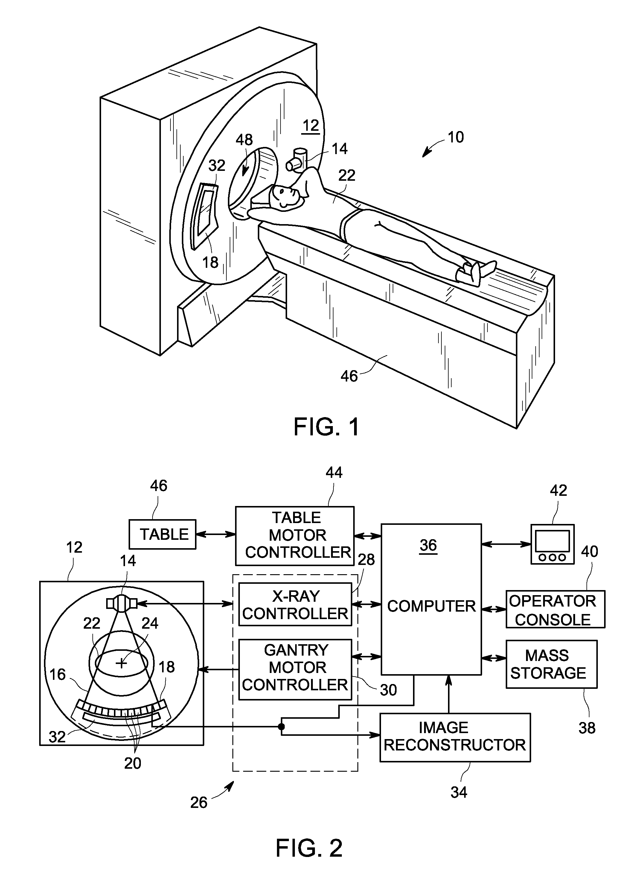
[0020]The operating environment of the invention is described with respect to a sixty-four-slice computed tomography (CT) system. However, it will be appreciated by those skilled in the art that the invention is equally applicable for use with single-slice or other multi-slice configurations. Moreover, the invention will be described with respect to the detection and conversion of x-rays. However, one skilled in the art will further appreciate that the invention is equally applicable for the detection and conversion of other high frequency electromagnetic energy. An implementation is employable with a “third generation” CT scanner and / or other CT systems.
[0021]Referring to FIG. 1, a computed tomography (CT) imaging system 10 is shown as including a gantry 12 representative of a “third generation” CT scanner. According to an exemplary embodiment of the invention, CT system 10 is provided as a Gemstone Spectral Imaging (GSI) dual energy CT system from GE Healthcare. Gantry 12 has an x...
PUM
 Abstract
Description
Claims
Application Information