
Application of nafamostat mesylate in the preparation of drugs for preventing and treating nervous system diseases
A technology of nafamostat mesylate and medicine, which is applied in the application field of nafamostat mesylate in the preparation of drugs for preventing and treating nervous system diseases
Patent Information
- Authority / Receiving Office
- CN · China
- Patent Type
- Patents(China)
- Current Assignee / Owner
- Publication Date
- 2018-07-17
Smart Images
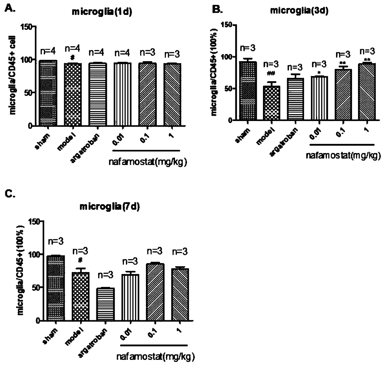
Figure 1 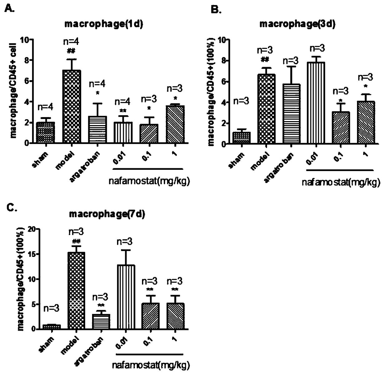
Figure 2 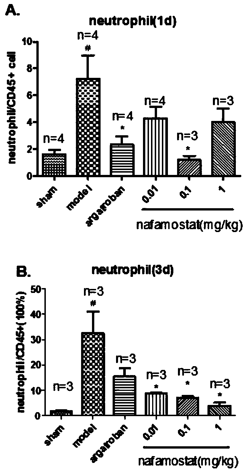
Figure 3
Abstract
Description
Technical field:
[0001] The invention belongs to the field of research and development of drugs for neurological diseases, and relates to a new drug application of nafamostat mesylate, in particular to the application of nafamostat mesylate in the preparation of drugs for preventing and treating neurological diseases. Background technique:
[0002] Nerve damage caused by ischemia, hypoxia, infection, trauma and other causes can cause corresponding neurological diseases such as cerebral ischemia (stroke), Alzheimer's disease (Alzheimer's disease, AD), epilepsy (epilepsy), Parkinson's disease Disease (Parkinson's disease, PD) and multiple sclerosis (multiple sclerosis, MS) and so on. These neurological diseases, especially cerebrovascular diseases, have seriously affected people's lives, and thus aroused widespread concern of scientists.
[0003] In the pathological process of many neurological diseases, neuroinflammation, blood-brain barrier damage, and neuronal injury and d...
Examples
Embodiment 1
[0022] Study on inflammatory cell migration of nafamostat mesylate in rat brain injury
[0023] 1. Experimental method
[0024] 1) Animal grouping
[0025] A total of 85 clean-grade male SD rats (certificate number: SCXK (Zhejiang) 2008-0033), weighing 250-280g. Sham operation group: 10; tMCAO model group: 15; positive drug group: 15; low-dose nafamostat mesilate group: 15; middle-dose nafamostat mesilate group: 15; Nafamostat sulfonate high dose group: 15 rats.
[0026] 2) Establishment of rat brain injury model (tMCAO)
[0027] Referring to the method of Longa et al., the rat tMCAO model was prepared by the internal carotid artery suture method: the rat was anesthetized with 3% chloral hydrate (300mg / kg, ip), fixed in supine position, routinely disinfected, and the neck was cut in the middle The skin was bluntly dissected in all layers, exposing the right CCA. After the ICA and the ECA bifurcation of the external carotid artery were separated, the vagus nerve and trache...
Embodiment 2
[0037] Study on the synthesis and release of inflammatory factors of nafamostat mesylate on brain injury in rats
[0038] 1. Experimental method
[0039] 1) Animal grouping
[0040] A total of 60 clean-grade male SD rats, weighing 250-280g. Sham operation group: 6; tMCAO model group: 12; positive drug group: 8; low-dose nafamostat mesilate group: 12; middle-dose nafamostat mesilate group: 12; Nafamostat sulfonate high-dose group: 12 rats.
[0041] 2) Establishment of rat brain injury model (tMCAO)
[0042] With embodiment 1.
[0043] 3) Method of administration
[0044] With embodiment 1.
[0045] 4) Index determination
[0046] After 22 hours of reperfusion, the rats were anesthetized with 3% chloral hydrate, and the heart was perfused with ice PBS. Immediately after taking the brain, it was frozen in liquid nitrogen.
[0047] ① Take an appropriate amount of cortical tissue from the brain lesion, extract mRNA, and perform qRT-PCR.
[0048] ②Take the cortical tissue ...
Embodiment 3
[0052] Effect of nafamostat mesylate on thrombin content and activity in rat brain after brain injury
[0053] 1. Experimental method
[0054] A total of 80 clean-grade male SD rats (certificate number: SCXK (Zhejiang) 2008-0033), weighing 250-280g. Sham operation group: 6; tMCAO model group: 15; positive drug group: 12; low-dose nafamostat mesilate group: 17; middle-dose nafamostat mesilate group: 15; Nafamostat sulfonate high dose group: 15 rats.
[0055] The model establishment and drug administration methods are the same as in Example 1. During the test, the animal is anesthetized, perfused with normal saline, and the brain is quickly taken out and placed on ice; sagittal slices are used to separate the left and right hemispheres; coronal slices of the damaged brain tissue are taken, 6-8 slices (The thickness of each slice is about 1 mm), and the striatum part of each slice is weighed separately. Transfer a part of the brain tissue into a doff tube, add 1 volume of PBS ...