
Methods for quantitating DNA using digital multiple displacement amplification
a technology of multiple displacement and quantitation method, applied in the field of methods for quantitating nucleic acids, can solve the problems of insufficient basis on which inability to distinguish composite genomes assembled from metagenomic data, and inability to accurately model the response of single-gene studies, etc., to reduce the impact of contaminates and reagent costs, improve the reliability and throughput of single-cell wga, and eliminate background amplification
Patent Information
- Authority / Receiving Office
- US · United States
- Patent Type
- Patents(United States)
- Current Assignee / Owner
- Publication Date
- 2019-11-26
Smart Images
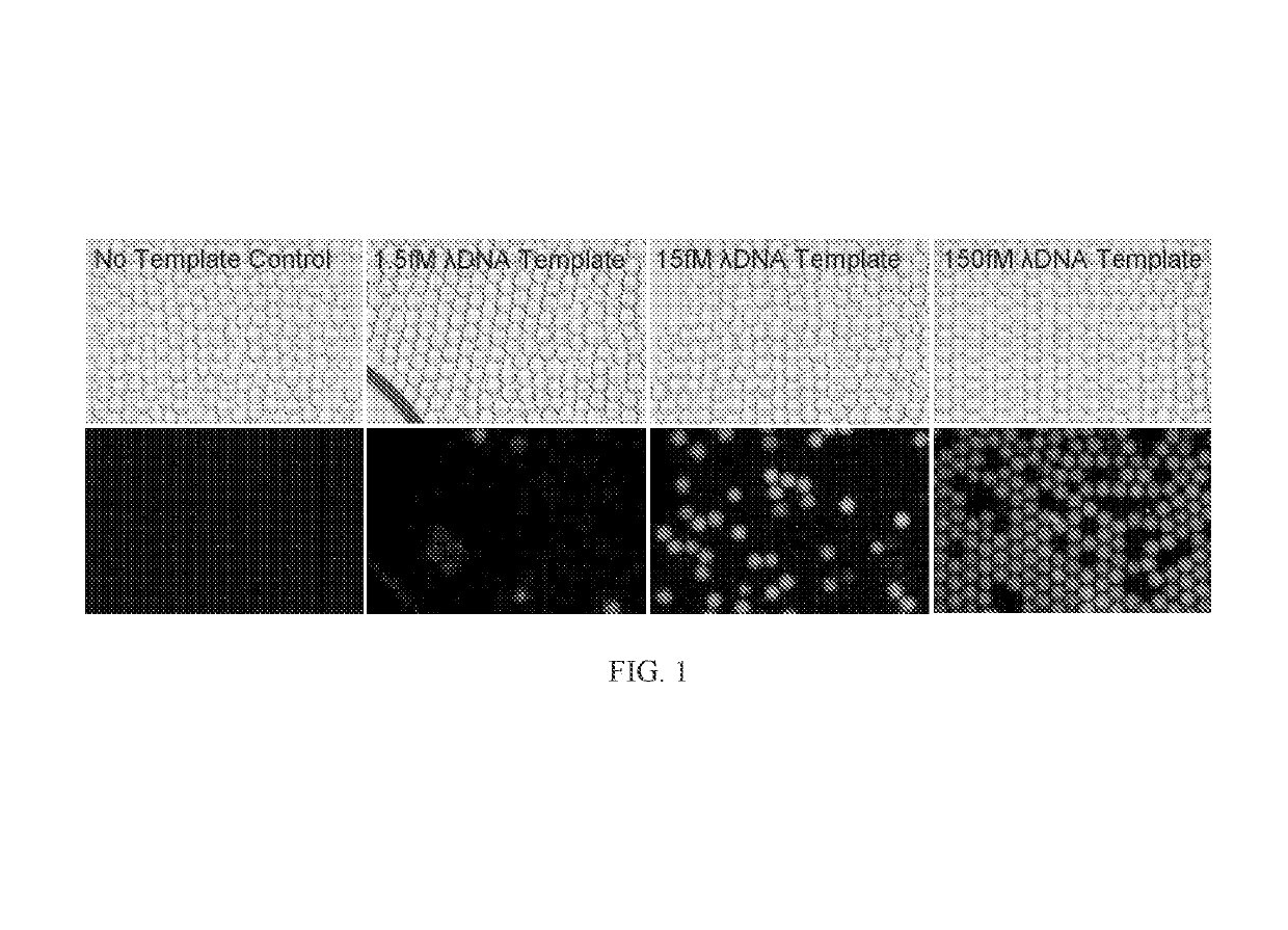
Figure 1 
Figure 2 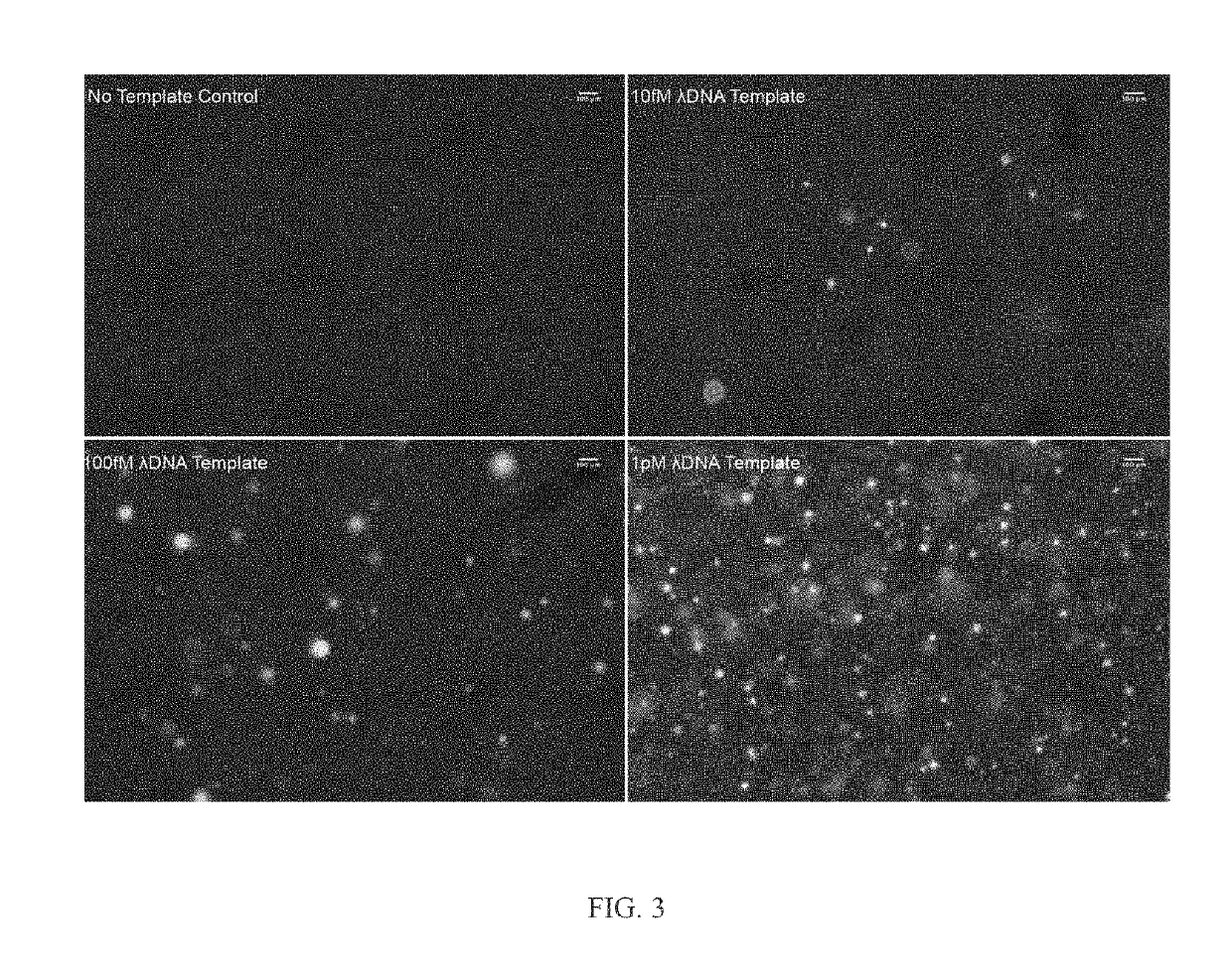
Figure 3
Abstract
Description
RELATED APPLICATIONS AND INCORPORATION BY REFERENCE
[0001] This application is a continuation-in-part of international patent application Serial No. PCT / US2014 / 028941, filed Mar. 14, 2014 and published as PCT Publication No. WO 2014 / 153071 on Sep. 23, 2014 and which claims benefit of and priority to U.S. provisional patent application Ser. No. 61 / 782,826 filed Mar. 14, 2013.FEDERAL FUNDING LEGEND
[0002] This invention was made with government support under Grant No. R01HG004863 awarded by the National Institutes of Health. The government has certain rights in the invention.
[0003] The foregoing application, and all documents cited therein or during their prosecution (“appln cited documents”) and documents cited or referenced herein (“herein cited documents”), and all documents cited or referenced in herein cited documents, together with any manufacturer's instructions, descriptions, product specifications, and product sheets for any products mentioned herein or in any document incorporate...
Examples
example 1
dic Droplets
[0095]Fabricating the Microfluidic Devices.
[0096]Microfluidic devices were constructed using standard PDMS soft lithography (McDonald J C, et al. (2000) Fabrication of microfluidic systems in poly(dimethylsilox-ane). Electrophoresis 21:27-40) with 18 micron SU8-on-Si wafer molds formed using transparency masks from FineLine Imaging. After curing, the PDMS device is adhered to glass for 2 hours at 80° C.
[0097]Multiple Displacement Reaction.
[0098]The MDA reactions were prepared according to protocol (Qiagen Repli-G Midi Kit) with Lambda DNA added to 5e-5, 5e-4, and 5e-3 ng / uL in the standard samples. Each reaction was supplemented with 0.3% Tween-20 and Evagreen prior to drop formation. Drop and bulk samples incubated at 30° C. for 16 h, and 65° C. for 20 m. Half of the post-incubation drop samples were converted to bulk with half volume 1H,1H,2H,2H-Perfluoro-1-octanol (Sigma Aldrich) and half of the post-incubation bulk samples were made into drops.
[0099]Forming Monodispe...
example 2
Matrices
[0108]FIGS. 3-6 depict MDA reactions in a gel matrix with lambda phage genome as a template, which is representative of single genome amplification as well as single cell analysis.
[0109]FIG. 3 depicts digital MDA products at 30° C. after 16 hours stained with 500 nM SYTOX Orange dye in 7% 4-Arm PEG Acrylate-Dithiol hydrogels. The initial Lambda Phage DNA template concentrations were 10 fM, 100 fM, and 1 pM. (or convert to mass / volume, 1.3e-5 ng / μL, 1.3e-4 ng / μL, and 1,3e-3 ng / μL).
[0110]FIG. 4 depicts a multiple displacement amplification in gel. The figure depicts the number of DNA amplification colonies observed vs. initial lambda phage DNA concentration in 4-arm PEG acrylate gel after 16 hours of multiple displacement amplification. DNA was stained with 500 nM SYTOX Orange.
[0111]FIG. 5 depicts 216 bp PCR products using Vent(exo-) polymerase (a polymerase with strong strand displacement) after 40 thermal cycles stained with 500 nM SYTOX Orange dye in 7% 4-Arm PEG Acrylate-D...
example 3
and Methods
[0113]4-Ami PEG Hydrogel. Hydrogel components, including 4-arm PEG acrylate (MW 10,000) and HS-PEG-SH (MW 3,400), were obtained from Layson Bio. For every 25 μl of 10% weight percentage cross-linked hydrogel, 1.6 mg of 4-arm PEG acrylate and 1.1 mg of HS-PEG-SH were dissolved in PBS buffer (Invitrogen). It was briefly vortexed and centrifuged to ensure mixing and it was allowed to sit for 10 minutes while the hydrogel components cross-linked through the reaction between the thiol and acrylate groups
[0114]Polymerase Chain Reaction (PCR). The primers used for PCR on purified λ DNA (48 kbp, NEB) were purchased from IDT. Primer F 5′: CGGCAAACGGGAATGAAACGCC. Primer R 5′: Alyson. A 25 μl hydrogel PCR reaction consisted of 2 U of VentR(exo-) polymerase (NEB), 1× ThermoPol Reaction Buffer (NEB), 0.4 mM dNTP (NEB), 1 μM Primers, 5% DMSO (Sigma), 0.5 mg / ml BSA(NEB), 1.6 mg 4-arm PEG acrylate in PBS, 1.1 mg HS-PEG-SH in PBS, and λDNA template (NEB) of various concentrations. The 25 ...