
Device to measure a radiation dose
Patent Information
- Authority / Receiving Office
- US · United States
- Current Assignee / Owner
- Publication Date
- 2007-06-05
- Estimated Expiration
- Not applicable · inactive patent
Smart Images
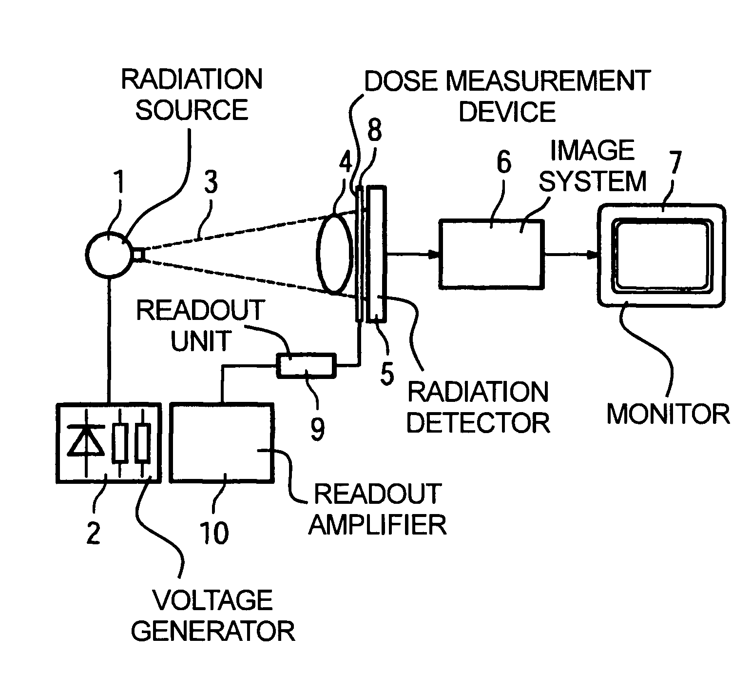
Figure 1 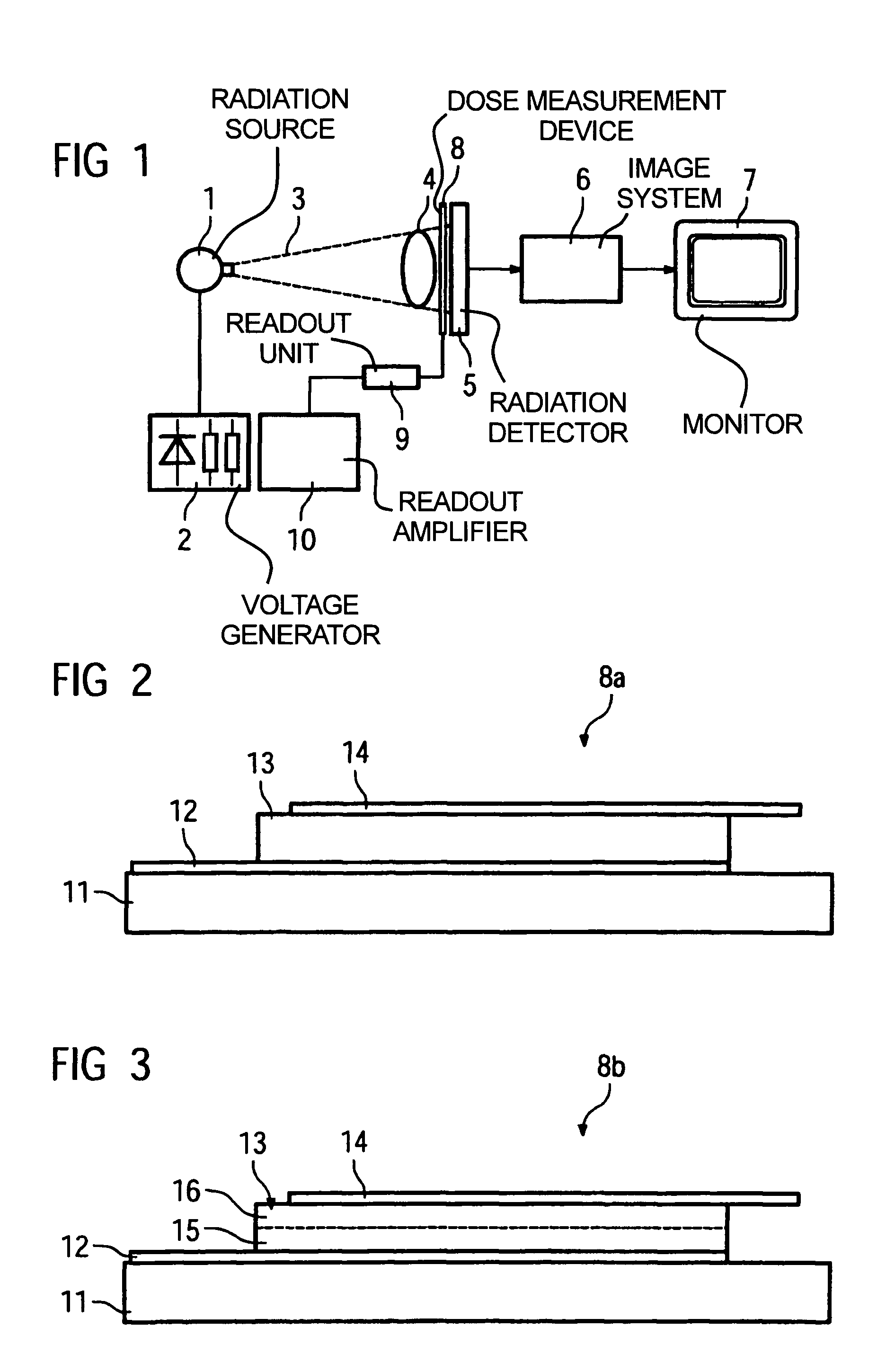
Figure 2 
Figure 3
Abstract
Description
BACKGROUND OF THE INVENTION
[0001] 1. Field of the Invention
[0002] The present invention concerns a device to measure a radiation dose, in particular an x-ray radiation dose, of the type that absorbs radiation and provides an output signal dependent on the absorption which represents a measure for the dose.
[0003] 2. Description of the Prior Art
[0004] In radiation image acquisition systems, in particular in x-ray systems as they are primarily used in medical diagnostics, it is necessary to regulate as precisely as possible the dose of the radiation that hits the patient to be examined. Typically an automatic dose regulator (Automatic Exposure Control) is used for this purpose, with either the radiation dose striking the detector or the unabsorbed radiation dose still present behind the detector being measured. The voltage generator of the x-ray tube is then regulated using this measurement value. Generally the duration of the emitted x-ray pulse is varied.
[0005] Conventionally, an ionizati...
Examples
Embodiment Construction
[0034]FIG. 1 shows an inventive radiation image acquisition apparatus having a radiation source 1, for example an x-ray tube with associated high-voltage generator 2 that generates an x-ray beam 3. After penetrating the subject or patient 4 to be examined, the radiation strikes on a detector 5, for example a solid-state planar detector based on amorphous silicon. The generated image signal is processed in an image system 6. The radiation images can be viewed on the monitor 7.
[0035]An inventive dose measurement device 8 that has a thin-film diode structure that provides a dose-dependent output signal is located in the beam path in front of the radiation detector 5. Downstream from the dose measurement device 8 is a readout unit 9 as well as a readout amplifier 10 via which the output signals of the dose measurement device 8 are read out and amplified, whereby the readout amplifier 10 then emits a control signal to the high-voltage generator 2 in order to therewith regulate the dose.
[...