Augmented lagrangian iterative reconstruction method of X-ray image and CI image

A CT image and iterative reconstruction technology, which is applied in the image processing field of medical imaging, can solve the problems that the objective function cannot be solved, the source of CT image noise is not considered, and the amount of calculation is large.

Inactive Publication Date: 2013-08-14
SOUTHERN MEDICAL UNIVERSITY
View PDF2 Cites 26 Cited by
  • Summary
  • Abstract
  • Description
  • Claims
  • Application Information

AI Technical Summary

Problems solved by technology

[0005] Strategy 1 does not consider the source of CT image noise, which belongs to post-processing technology. The main problem is that the distribution characteristics of CT image noise are complex, and it is difficult to implement high-performance filters.
Non-smooth regularization terms in iterative reconstruction of CT images, such as total variation and l 1 Regularization, so that the corresponding objective function cannot be solved by the traditional gradient descent (Gradient Descend) method
Although a smooth regularization function can be used for approximation, the corresponding algorithm is computationally intensive and slow to converge
For the smooth regularization in iterative reconstruction of CT images, although it can be solved by traditional gradient descent methods, such as the nonlinear conjugate gradient method (NCG), the calculation amount is often large and the convergence speed is slow

Method used


Image

  • Augmented lagrangian iterative reconstruction method of X-ray image and CI image
  • Augmented lagrangian iterative reconstruction method of X-ray image and CI image
  • Augmented lagrangian iterative reconstruction method of X-ray image and CI image

Examples


Embodiment 1

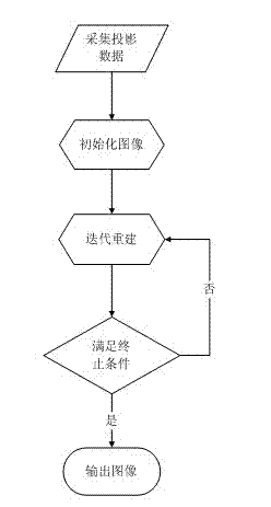
[0064] An augmented Lagrangian iterative reconstruction method of an X-ray CT image, comprising the steps in turn:

[0065] (1) Obtain the system parameters of the CT equipment and the projection data y under the low-dose scanning protocol; the acquired system parameters of the CT equipment mainly include the X-ray incident photon intensity I 0 and the variance of the electronic noise of the system Wait.

[0066] (2) For the projection data y in step (1), the variance on each data point is performed estimate, and use the FBP method to reconstruct the projection data y in step (1) to obtain the initial image.

[0067] (3) Perform iterative reconstruction using the initial image obtained in step (2) as the iterative initial image to obtain a final reconstructed image.

[0068] The iterative reconstruction in step (3) is carried out by the following method,

[0069] Iterative reconstruction aims at the following X-ray CT image reconstruction model:

[0070] ...

Embodiment 2

[0086] An augmented Lagrangian iterative reconstruction method for an X-ray CT image, comprising the following steps in turn: (1) Obtaining system parameters of a CT device and projection data y under a low-dose scanning protocol; obtaining system parameters of the CT device Mainly includes X-ray incident photon intensity I 0 and the variance of the electronic noise of the system Wait.

[0087] (2) For the projection data y in step (1), the variance on each data point is performed estimate, and use the FBP method to reconstruct the projection data y in step (1) to obtain the initial image.

[0088] (3) Using the initial image obtained in step (2) as an iterative initial image to perform iterative reconstruction to obtain a final reconstructed image.

[0089] The iterative reconstruction in step (3) is carried out by the following method,

[0090] Iterative reconstruction aims at the following X-ray CT image reconstruction model:

[0091] ...

Embodiment 3

[0125] An augmented Lagrangian iterative reconstruction method of an X-ray CT image, other contents are the same as in embodiment 2, the difference is: for the following non-smooth objective function, the formula (5) is solved, specifically as follows:

[0126] For formula (5), we can solve it exactly. (5) can be written as multiple one-dimensional minimization problems:

[0127] x k + 1 r = arg min x r Ψ ( x r ) + βλ k r 2 ( x r - ρ k r ...


PUM


Abstract

An augmented lagrangian iterative reconstruction method of an X-ray image and a CI image is characterized by comprising the following steps: (1) acquiring system parameters and projecting data under a low dose scanning protocol of CI equipment; (2) carrying out one-by-one data point variance estimation on the projecting data in the step (1), and conducting filtered back projection on the projected data in the step (1) to obtain an initial pattern; (3) taking the initial pattern obtained from the step (2) as an iterative initial pattern to be subjected to iterative reconstruction, and obtaining the final reconstruction pattern through loop iteration according to the iterative formula. The invention provides an algorithm for optimizing the iterative formula. The method has the advantages of wide application range, less iterative times, and high imaging quality.

Description

technical field [0001] The invention relates to the technical field of image processing of medical images, in particular to an augmented Lagrangian iterative reconstruction method of X-ray CT images. Background technique [0002] X-ray CT scanning has been widely used in clinical medical imaging diagnosis, but X-ray radiation dose (Radiation Dose) will cause radiation damage to the human body, and even induce unpredictable damage such as malignant tumors. In order to reduce the damage to the user, it has become an urgent requirement in the field of medical CT imaging to minimize the X-ray dose on the premise of ensuring the quality of the reconstructed image. [0003] In order to reduce the dose of X-ray radiation, various hardware and software technologies can be used to reduce the dose of X-ray used in CT scanning. Common methods include reducing tube current, reducing X-ray exposure time, and reducing the acquisition angle of projection data. However, low-dose (Low Dose...

Claims


Application Information

Patent Timeline
Patent Type & Authority Applications(China)
IPC IPC(8): G06T11/00
Inventor 马建华牛善洲黄静陈武凡
Owner SOUTHERN MEDICAL UNIVERSITY
Why Eureka
Try Eureka