Ultrasonic medical device, and ultrasonic image diagnostic device

A medical device, ultrasonic technology, applied in ultrasonic/sonic/infrasonic diagnosis, sonic diagnosis, infrasound diagnosis, etc., can solve the problem of inability to supply liquid food through the mouth, etc.

Active Publication Date: 2014-09-24
TOSHIBA MEDICAL SYST CORP
View PDF7 Cites 9 Cited by
  • Summary
  • Abstract
  • Description
  • Claims
  • Application Information

AI Technical Summary

Problems solved by technology

[0013] However, if the insertion part is closely attached to the inner wall of the esophagus, there is a problem that liquid food or the like cannot pass through the esophagus of the subject orally.

Method used


Image

  • Ultrasonic medical device, and ultrasonic image diagnostic device
  • Ultrasonic medical device, and ultrasonic image diagnostic device
  • Ultrasonic medical device, and ultrasonic image diagnostic device

Examples


no. 1 Embodiment approach

[0045] A first embodiment of the ultrasonic imaging diagnostic apparatus will be described with reference to the drawings.

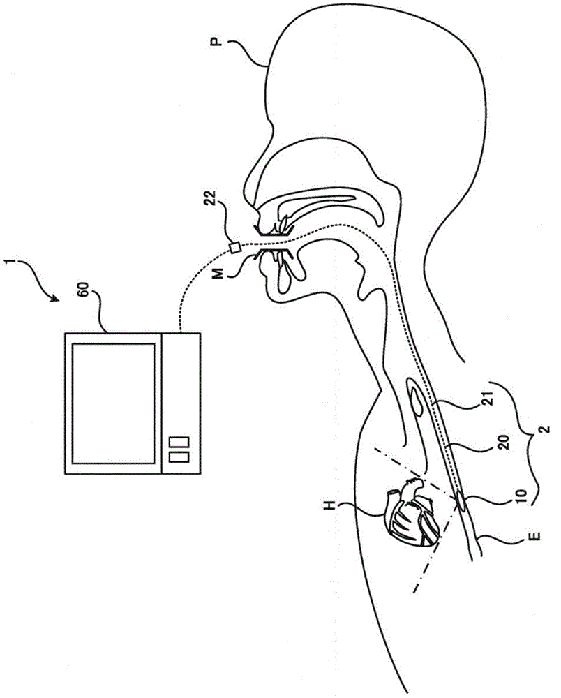
[0046] figure 1 An example is shown in , in which the ultrasonic imaging diagnostic apparatus 1 according to this embodiment indwells the capsule body part 10 at a desired position in the esophagus E, and indwells the desired position of the subject P in the indwelling state. An organ (heart H) transmits ultrasonic waves, receives reflected waves from the heart H as echo signals, and observes the heart H. Hereinafter, the transmission of ultrasonic waves and the reception of reflected waves may be collectively referred to as "transmission and reception of ultrasonic waves". The capsule body unit 10 transmits echo signals to the external device 60 , and the external device 60 processes the signal received from the capsule body unit 10 to create and display an ultrasonic image. In addition, the heart H shown in each drawing is schematically shown in orde...

no. 2 Embodiment approach

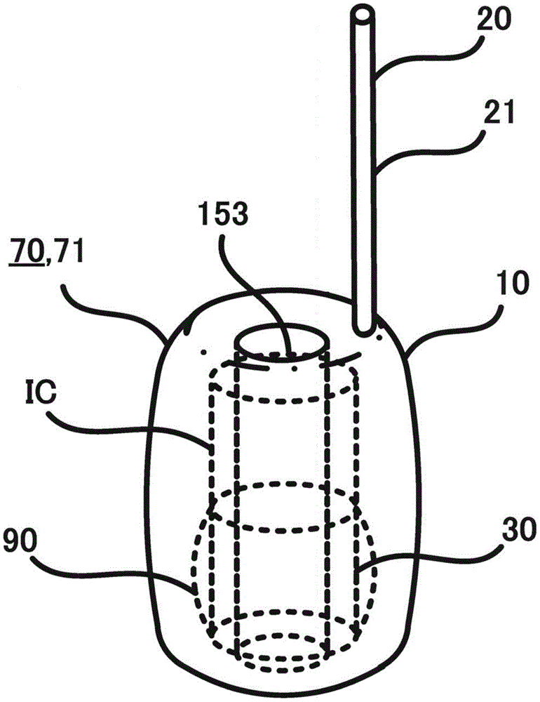
[0134] Next, refer to Figure 14 as well as Figure 15 , and a second embodiment of the ultrasonic medical device will be described. Figure 14 It is a longitudinal sectional view of the capsule-shaped main body when the expansion-contraction body 70 is inflated, Figure 15 It is a vertical cross-sectional view of the capsule-shaped main body when the expandable-contractable body 70 is contracted.

[0135] In addition, in 2nd Embodiment, the same code|symbol is attached|subjected to the same structure as 1st Embodiment, and description is abbreviate|omitted, and a different structure is mainly demonstrated.

[0136] like Figure 14 as well as Figure 15 As shown, the configurations of the support 15 , the ultrasonic vibrator 30 , the integrated circuit IC, the ultrasonic motor 42 , and the acoustic lens 90 are the same as those of the first embodiment.

[0137] In the first embodiment, only the expandable body 70 that expands and contracts the bag-shaped container 71 is s...

no. 3 Embodiment approach

[0147] Next, refer to Figure 16 A third embodiment of the ultrasonic medical device will be described. Figure 16 It is a cross-sectional view of the capsule-shaped main body when the expansion-contraction body is contracted.

[0148] In addition, in the third embodiment, the same reference numerals are assigned to the same configurations as those in the first embodiment, and description thereof will be omitted, and different configurations will be mainly described.

[0149] In the first embodiment, only the expandable body 70 that expands and contracts the bag-shaped container 71 is shown. However, it is not limited thereto, and in the second embodiment, the bag-shaped container 71 that is folded when contracted and unfolded when expanded is used.

[0150] like Figure 16 As shown, accordion-shaped creases are provided on the peripheral surface (full circumference) of the bag-shaped container 71 . By filling the contracted bag-shaped container 71 with a liquid or the lik...


PUM


Abstract

Provided are an ultrasonic image diagnostic device, and an ultrasonic medical device which can orally pass liquid food and the like through the esophagus of a subject. The ultrasonic medical device of an embodiment has a capsule-shaped main body part having an in-built ultrasonic transducer, transmits ultrasonic waves to the inside of the subject from the ultrasonic transducer inside the capsule-shaped main body after the same has been inserted into a tubular portion of the subject, and then receives the reflected waves. The ultrasonic medical device also has a support body. The support body is built into the capsule-shaped main body part and has a cylindrical shape. A plurality of ultrasonic transducers are arranged on the outer part of the cylinder, and a through hole is provided that penetrates the cylinder in the axial direction thereof.

Description

technical field [0001] Embodiments of the present invention relate to an ultrasonic medical device and an ultrasonic image diagnosis device. Background technique [0002] Ultrasonic imaging diagnostic devices use an ultrasonic probe to scan the inside of the subject with ultrasonic waves, and image the inside of the subject based on data generated from the reflected waves. [0003] An example of an ultrasonic probe used in an ultrasonic imaging diagnostic apparatus includes a TEE (trans-esophageal echocardiogram) probe (for example, Patent Document 1). The TEE probe is inserted into the esophagus, for example, through the mouth, and is used for imaging the heart and the like. The TEE probe has a central tube inserted into the esophagus, and an insertion portion provided at the distal end of the central tube. [0004] A power line for transmitting power to the ultrasonic vibrators, a control line for transmitting control signals to the ultrasonic vibrators, and a data line ...

Claims


Application Information

Patent Timeline
Patent Type & Authority Applications(China)
IPC IPC(8): A61B8/12
CPCA61B1/041G01S7/5208B06B2201/76A61B8/0883A61B8/445B06B1/0215A61B8/12A61B8/4494B06B1/0633
Inventor 山形仁平间信
Owner TOSHIBA MEDICAL SYST CORP
Why Eureka
Try Eureka